Sergey V. Petryakov, Maciej M. Kmiec, Ryan C. O’Connell, Conner S. Ubert, Victor B. Kassey, Philip E. Schaner, Periannan Kuppusamy
{"title":"A Cylindrical Surface Dielectric Resonator with Substantially High Sensitivity for Deep-Tissue EPR Oximetry","authors":"Sergey V. Petryakov, Maciej M. Kmiec, Ryan C. O’Connell, Conner S. Ubert, Victor B. Kassey, Philip E. Schaner, Periannan Kuppusamy","doi":"10.1007/s00723-024-01747-8","DOIUrl":null,"url":null,"abstract":"<div><p>Electron paramagnetic resonance (EPR) has been established as a unique and reliable method for quantitative in vivo oximetry applicable to a variety of preclinical and clinical studies. A recent clinical study using EPR oximetry with OxyChip from our laboratory demonstrated the feasibility of tumor oxygen measurements in cancer patients (Schaner, et al. Front. Oncol. 2021). During this study, the need to improve oxygen measurement capability in tumors at depths greater than 10 mm became apparent. This prompted us to develop new designs of resonators (RF coils) with enhanced sensitivity for measuring deep-tissue oxygen levels. In this manuscript, we report the development of a new cylindrical surface dielectric resonator (c-SDR) designed with a ceramic dielectric material for substantially enhanced sensitivity and capability for deep-tissue oximetry. The c-SDR was constructed with a cylindrical dielectric material (<i>ϕ</i> 27.2 × 22.2 mm; <i>ε</i> = 160), 6-segmented coupling loop and copper shield to provide an active surface (aperture) of 25 mm with an operating frequency of 1.16 GHz (L-band) and an unloaded <i>Q</i> 600. The resonator could detect OxyChip (<i>ϕ</i> 0.6 × 5 mm) at a surface-to-sample depth of 50 mm in water or 30 mm in a tissue-emulating phantom with a signal-to-noise ratio of 5. Further evaluations of the c-SDR using OxyChip demonstrated its capability for oxygen measurements at depths of 27 mm for 1% oxygen and 15 mm for 5% oxygen in a tissue phantom. In conclusion, the new c-SDR is a significant upgrade to the currently used resonators for in vivo EPR oximetry including clinical oximetry.</p></div>","PeriodicalId":469,"journal":{"name":"Applied Magnetic Resonance","volume":"56 5","pages":"613 - 629"},"PeriodicalIF":1.1000,"publicationDate":"2024-12-31","publicationTypes":"Journal Article","fieldsOfStudy":null,"isOpenAccess":false,"openAccessPdf":"","citationCount":"0","resultStr":null,"platform":"Semanticscholar","paperid":null,"PeriodicalName":"Applied Magnetic Resonance","FirstCategoryId":"101","ListUrlMain":"https://link.springer.com/article/10.1007/s00723-024-01747-8","RegionNum":4,"RegionCategory":"物理与天体物理","ArticlePicture":[],"TitleCN":null,"AbstractTextCN":null,"PMCID":null,"EPubDate":"","PubModel":"","JCR":"Q4","JCRName":"PHYSICS, ATOMIC, MOLECULAR & CHEMICAL","Score":null,"Total":0}
引用次数: 0
Abstract
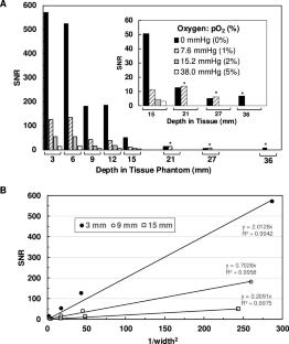
Electron paramagnetic resonance (EPR) has been established as a unique and reliable method for quantitative in vivo oximetry applicable to a variety of preclinical and clinical studies. A recent clinical study using EPR oximetry with OxyChip from our laboratory demonstrated the feasibility of tumor oxygen measurements in cancer patients (Schaner, et al. Front. Oncol. 2021). During this study, the need to improve oxygen measurement capability in tumors at depths greater than 10 mm became apparent. This prompted us to develop new designs of resonators (RF coils) with enhanced sensitivity for measuring deep-tissue oxygen levels. In this manuscript, we report the development of a new cylindrical surface dielectric resonator (c-SDR) designed with a ceramic dielectric material for substantially enhanced sensitivity and capability for deep-tissue oximetry. The c-SDR was constructed with a cylindrical dielectric material (ϕ 27.2 × 22.2 mm; ε = 160), 6-segmented coupling loop and copper shield to provide an active surface (aperture) of 25 mm with an operating frequency of 1.16 GHz (L-band) and an unloaded Q 600. The resonator could detect OxyChip (ϕ 0.6 × 5 mm) at a surface-to-sample depth of 50 mm in water or 30 mm in a tissue-emulating phantom with a signal-to-noise ratio of 5. Further evaluations of the c-SDR using OxyChip demonstrated its capability for oxygen measurements at depths of 27 mm for 1% oxygen and 15 mm for 5% oxygen in a tissue phantom. In conclusion, the new c-SDR is a significant upgrade to the currently used resonators for in vivo EPR oximetry including clinical oximetry.
期刊介绍:
Applied Magnetic Resonance provides an international forum for the application of magnetic resonance in physics, chemistry, biology, medicine, geochemistry, ecology, engineering, and related fields.
The contents include articles with a strong emphasis on new applications, and on new experimental methods. Additional features include book reviews and Letters to the Editor.
 求助内容:
求助内容: 应助结果提醒方式:


