{"title":"Percutaneous nephrolithotomy: wisdom, dogma, paradigm and myths surrounding puncture","authors":"Peter Alken","doi":"10.1111/bju.16740","DOIUrl":null,"url":null,"abstract":"<p>A non-transpapillary technique [<span>1</span>] seems to facilitate access to the kidney, the most crucial aspect of percutaneous nephrolithotomy, compared to the classic transpapillary method [<span>2</span>].</p>\n<p>In modern times, the godfathers of percutaneous access, Goodwin et al. [<span>3</span>], who illustrated a pyelostomy rather than a nephrostomy in Fig. 2 of their 1955 article, should be commended for their non-papillary puncture.</p>\n<p>From the contemporary first accounts of ‘experience with a central, noncalyceal puncture protocol for percutaneous nephrolithotripsy’ [<span>4</span>] in 2017, a myth has persisted that has carried through to all subsequent studies on the subject, including the study by Lotfi et al. [<span>1</span>], that the ‘Current understanding of anatomical background of percutaneous access is based mostly on the very extensive documentation by Sampaio’ [<span>4</span>].</p>\n<p>By the time of Sampaio's publications, 15 years after the introduction of endoscopic PNL in the late 1970s, several thousand PNLs had probably been performed worldwide based on the transpapillary principle. The first instruments specifically designed in 1980 for endoscopically controlled PNL [<span>2</span>] followed two principles: Access through the least vascularised part of the parenchyma and access with the nephrosocpe into the collecting system at the point where it is connected to the parenchyma, i.e. the collecting system itself should not be injured. The aim was to avoid vascular trauma and extravasation and also to reach even the most peripherally located calyceal stone.</p>\n<p>This was the reversal of the least traumatic way to place a nephrostomy tube into the collecting system from the point of view of a urologist influenced by having previously performed open surgery. In the times of open surgery, a forceps was pushed transpapillary from the calyx, an anatomically preformed tract, to the surface of the kidney in order to pull the nephrostomy tube into the collecting system. In the late 1960s, John Wickham [<span>5</span>] added the open transpapillary avascular multiple radial nephrotomies technique for the removal of staghorn calculi to this decades-old transpapillary technique. This was later refined by a team at the University of Mainz, who performed staghorn surgery without clamping the renal artery, utilising only transpapillary access.</p>\n<p>In PNL, I have not always hit the mark with a perfect transpapillary approach when carrying out PNL, but was sometimes happy simply to obtain access (Fig. 1). In one of his many articles on open stone surgery, Wickham described ‘Large venous anastomoses … like collars around the calyceal necks.’ In my experience and that of others (Tursunkulov AN, Akfamedline University Hospital, Central Asian University, Tashkent; personal communication), this description fits the annoying venous oozing frequently observed behind the nephroscope when using the non-papillary access method, which can usually be stopped temporarily by slightly angling the instrument and thus compressing the open veins. As long as it is of venous origin, this is only a temporary problem.</p>\n<figure><picture>\n<source media=\"(min-width: 1650px)\" srcset=\"/cms/asset/6cb02b15-0fa7-42b5-95b8-6f9ff949fd01/bju16740-fig-0001-m.jpg\"/><img alt=\"Details are in the caption following the image\" data-lg-src=\"/cms/asset/6cb02b15-0fa7-42b5-95b8-6f9ff949fd01/bju16740-fig-0001-m.jpg\" loading=\"lazy\" src=\"/cms/asset/f732455e-47b9-4237-8561-9a1d1e0dcce2/bju16740-fig-0001-m.png\" title=\"Details are in the caption following the image\"/></picture><figcaption>\n<div><strong>Fig. 1<span style=\"font-weight:normal\"></span></strong><div>Open in figure viewer<i aria-hidden=\"true\"></i><span>PowerPoint</span></div>\n</div>\n<div>‘Happy to obtain access’ despite a transpyelic approach with subsequent renal displacement due to a mixed urinoma haematoma caused by an artery lesion in the renal hilum.</div>\n</figcaption>\n</figure>\n<p>Otherwise, ‘Happy to obtain access’ seems to be a PNL rule, as many others have already indirectly stated. For example, Tahra et al. reported that ‘we puncture wherever we can to achieve stone-free status and reduce unnecessary access…’ in their 11-year experience of performing PNLs using non-papillary access in 207 patients and papillary access in 69 patients [<span>6</span>]. Or, as Cracco and Scoffone wisely stated: ‘Consider also that endourologists routinely performing the papillary puncture for PCNL for sure will not carry out perfect punctures in 100% of the cases, therefore the current literature actually includes the outcomes of thousands of both ‘real papillary’ and ‘semi-papillary’ punctures’ [<span>7</span>]. The authors of the present study concluded [<span>1</span>]: ‘The non-papillary approach could be considered in the context of a different viable access gaining technique, especially when papillary access is unfeasible or technically challenging.’</p>\n<p>However, I am still not happy with ‘Targeting a larger area (calyces, infundibulums and pelvis) than a single point (tip of the calyx)’ [<span>4</span>]. The catastrophe does not have to occur frequently, but it can have a significant impact on the individual patient and the surgeon. Sampaio, who stated in 1988, based not on clinical but on experimental studies, ‘During endourological renal stone removal one of the most neglected aspects is that of anatomy’ [<span>8</span>], should also be requested to comment on the ongoing debate on the theory and practice of anatomy.</p>","PeriodicalId":8985,"journal":{"name":"BJU International","volume":"136 1","pages":""},"PeriodicalIF":3.7000,"publicationDate":"2025-04-15","publicationTypes":"Journal Article","fieldsOfStudy":null,"isOpenAccess":false,"openAccessPdf":"","citationCount":"0","resultStr":null,"platform":"Semanticscholar","paperid":null,"PeriodicalName":"BJU International","FirstCategoryId":"3","ListUrlMain":"https://doi.org/10.1111/bju.16740","RegionNum":2,"RegionCategory":"医学","ArticlePicture":[],"TitleCN":null,"AbstractTextCN":null,"PMCID":null,"EPubDate":"","PubModel":"","JCR":"Q1","JCRName":"UROLOGY & NEPHROLOGY","Score":null,"Total":0}
引用次数: 0
Abstract
A non-transpapillary technique [1] seems to facilitate access to the kidney, the most crucial aspect of percutaneous nephrolithotomy, compared to the classic transpapillary method [2].
In modern times, the godfathers of percutaneous access, Goodwin et al. [3], who illustrated a pyelostomy rather than a nephrostomy in Fig. 2 of their 1955 article, should be commended for their non-papillary puncture.
From the contemporary first accounts of ‘experience with a central, noncalyceal puncture protocol for percutaneous nephrolithotripsy’ [4] in 2017, a myth has persisted that has carried through to all subsequent studies on the subject, including the study by Lotfi et al. [1], that the ‘Current understanding of anatomical background of percutaneous access is based mostly on the very extensive documentation by Sampaio’ [4].
By the time of Sampaio's publications, 15 years after the introduction of endoscopic PNL in the late 1970s, several thousand PNLs had probably been performed worldwide based on the transpapillary principle. The first instruments specifically designed in 1980 for endoscopically controlled PNL [2] followed two principles: Access through the least vascularised part of the parenchyma and access with the nephrosocpe into the collecting system at the point where it is connected to the parenchyma, i.e. the collecting system itself should not be injured. The aim was to avoid vascular trauma and extravasation and also to reach even the most peripherally located calyceal stone.
This was the reversal of the least traumatic way to place a nephrostomy tube into the collecting system from the point of view of a urologist influenced by having previously performed open surgery. In the times of open surgery, a forceps was pushed transpapillary from the calyx, an anatomically preformed tract, to the surface of the kidney in order to pull the nephrostomy tube into the collecting system. In the late 1960s, John Wickham [5] added the open transpapillary avascular multiple radial nephrotomies technique for the removal of staghorn calculi to this decades-old transpapillary technique. This was later refined by a team at the University of Mainz, who performed staghorn surgery without clamping the renal artery, utilising only transpapillary access.
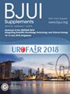
In PNL, I have not always hit the mark with a perfect transpapillary approach when carrying out PNL, but was sometimes happy simply to obtain access (Fig. 1). In one of his many articles on open stone surgery, Wickham described ‘Large venous anastomoses … like collars around the calyceal necks.’ In my experience and that of others (Tursunkulov AN, Akfamedline University Hospital, Central Asian University, Tashkent; personal communication), this description fits the annoying venous oozing frequently observed behind the nephroscope when using the non-papillary access method, which can usually be stopped temporarily by slightly angling the instrument and thus compressing the open veins. As long as it is of venous origin, this is only a temporary problem.
Fig. 1
Open in figure viewerPowerPoint
‘Happy to obtain access’ despite a transpyelic approach with subsequent renal displacement due to a mixed urinoma haematoma caused by an artery lesion in the renal hilum.
Otherwise, ‘Happy to obtain access’ seems to be a PNL rule, as many others have already indirectly stated. For example, Tahra et al. reported that ‘we puncture wherever we can to achieve stone-free status and reduce unnecessary access…’ in their 11-year experience of performing PNLs using non-papillary access in 207 patients and papillary access in 69 patients [6]. Or, as Cracco and Scoffone wisely stated: ‘Consider also that endourologists routinely performing the papillary puncture for PCNL for sure will not carry out perfect punctures in 100% of the cases, therefore the current literature actually includes the outcomes of thousands of both ‘real papillary’ and ‘semi-papillary’ punctures’ [7]. The authors of the present study concluded [1]: ‘The non-papillary approach could be considered in the context of a different viable access gaining technique, especially when papillary access is unfeasible or technically challenging.’
However, I am still not happy with ‘Targeting a larger area (calyces, infundibulums and pelvis) than a single point (tip of the calyx)’ [4]. The catastrophe does not have to occur frequently, but it can have a significant impact on the individual patient and the surgeon. Sampaio, who stated in 1988, based not on clinical but on experimental studies, ‘During endourological renal stone removal one of the most neglected aspects is that of anatomy’ [8], should also be requested to comment on the ongoing debate on the theory and practice of anatomy.
期刊介绍:
BJUI is one of the most highly respected medical journals in the world, with a truly international range of published papers and appeal. Every issue gives invaluable practical information in the form of original articles, reviews, comments, surgical education articles, and translational science articles in the field of urology. BJUI employs topical sections, and is in full colour, making it easier to browse or search for something specific.

 求助内容:
求助内容: 应助结果提醒方式:


