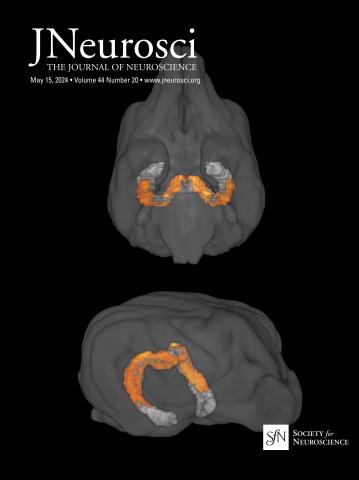
Multimodal Correspondence Between Optogenetic fMRI, Electrophysiology, and Anatomical Maps of the Secondary Somatosensory Cortex in Nonhuman Primates.

IF 4.4 2区 医学 Q1 NEUROSCIENCES
Pai-Feng Yang,Jamie Reed,Zhangyan Yang,Feng Wang,Ning Zheng,John C Gore,Li Min Chen
{"title":"Multimodal Correspondence Between Optogenetic fMRI, Electrophysiology, and Anatomical Maps of the Secondary Somatosensory Cortex in Nonhuman Primates.","authors":"Pai-Feng Yang,Jamie Reed,Zhangyan Yang,Feng Wang,Ning Zheng,John C Gore,Li Min Chen","doi":"10.1523/jneurosci.2375-24.2025","DOIUrl":null,"url":null,"abstract":"Optogenetic neuromodulation combined with fMRI (opto-fMRI) enables noninvasive monitoring of brain-wide activity and probs causal connections. In this study, we focused on the secondary somatosensory cortex (S2), a hub for integrating tactile and nociceptive information. By selectively stimulating excitatory neurons in the S2 cortex of monkeys using optogenetics, we observed widespread opto-fMRI activity in regions beyond the somatosensory system, as well as a strong spatial correspondence between opto-fMRI activity map and anatomical connections of the S2 cortex. Locally, optogenetically evoked fMRI BOLD signals from putative excitatory neurons exhibited standard hemodynamic response function. At low laser power, graded opto-fMRI signal changes are closely correlated with increases in LFP signals, but not with spiking activity. This indicates that LFP changes in excitatory neurons more accurately reflect the opto-fMRI signals than spikes. In summary, our optogenetic fMRI and anatomical findings provide causal functional and anatomical evidence supporting the role of the S2 cortex as a critical hub connecting sensory regions to higher-order cortical and subcortical regions involved in cognition and emotion. The electrophysiological basis of the opto-fMRI signals uncovered in this study offers novel insights into interpreting opto-fMRI results. Non-human primates are an invaluable intermediate model for translating optogenetic preclinical findings to humans.Significance statement The neocortex consists of excitatory and inhibitory neurons, each playing distinct roles in cognition and contributing to brain disorders. This study marks the first to use optogenetics to selectively stimulate excitatory neurons of the primate somatosensory S2 cortex, establishing a spatial correspondence between brain-wide opto-fMRI activity and anatomical tracer-based connections in the S2 hand region. The differential relationships between laser power and changes in BOLD, local field potentials, and spiking activity suggest that LFP signals better reflect opto-fMRI signals at the excitatory neuron population level. This work has translational potential for developing targeted neuromodulation therapies for neurological and psychiatric disorders. Success in NHPs, along with advances in noninvasive AAV delivery, paves the way for future clinical applications.","PeriodicalId":50114,"journal":{"name":"Journal of Neuroscience","volume":"25 1","pages":""},"PeriodicalIF":4.4000,"publicationDate":"2025-04-09","publicationTypes":"Journal Article","fieldsOfStudy":null,"isOpenAccess":false,"openAccessPdf":"","citationCount":"0","resultStr":null,"platform":"Semanticscholar","paperid":null,"PeriodicalName":"Journal of Neuroscience","FirstCategoryId":"3","ListUrlMain":"https://doi.org/10.1523/jneurosci.2375-24.2025","RegionNum":2,"RegionCategory":"医学","ArticlePicture":[],"TitleCN":null,"AbstractTextCN":null,"PMCID":null,"EPubDate":"","PubModel":"","JCR":"Q1","JCRName":"NEUROSCIENCES","Score":null,"Total":0}
引用次数: 0

Abstract

Optogenetic neuromodulation combined with fMRI (opto-fMRI) enables noninvasive monitoring of brain-wide activity and probs causal connections. In this study, we focused on the secondary somatosensory cortex (S2), a hub for integrating tactile and nociceptive information. By selectively stimulating excitatory neurons in the S2 cortex of monkeys using optogenetics, we observed widespread opto-fMRI activity in regions beyond the somatosensory system, as well as a strong spatial correspondence between opto-fMRI activity map and anatomical connections of the S2 cortex. Locally, optogenetically evoked fMRI BOLD signals from putative excitatory neurons exhibited standard hemodynamic response function. At low laser power, graded opto-fMRI signal changes are closely correlated with increases in LFP signals, but not with spiking activity. This indicates that LFP changes in excitatory neurons more accurately reflect the opto-fMRI signals than spikes. In summary, our optogenetic fMRI and anatomical findings provide causal functional and anatomical evidence supporting the role of the S2 cortex as a critical hub connecting sensory regions to higher-order cortical and subcortical regions involved in cognition and emotion. The electrophysiological basis of the opto-fMRI signals uncovered in this study offers novel insights into interpreting opto-fMRI results. Non-human primates are an invaluable intermediate model for translating optogenetic preclinical findings to humans.Significance statement The neocortex consists of excitatory and inhibitory neurons, each playing distinct roles in cognition and contributing to brain disorders. This study marks the first to use optogenetics to selectively stimulate excitatory neurons of the primate somatosensory S2 cortex, establishing a spatial correspondence between brain-wide opto-fMRI activity and anatomical tracer-based connections in the S2 hand region. The differential relationships between laser power and changes in BOLD, local field potentials, and spiking activity suggest that LFP signals better reflect opto-fMRI signals at the excitatory neuron population level. This work has translational potential for developing targeted neuromodulation therapies for neurological and psychiatric disorders. Success in NHPs, along with advances in noninvasive AAV delivery, paves the way for future clinical applications.
求助全文
约1分钟内获得全文 求助全文
来源期刊
Journal of Neuroscience
Journal of Neuroscience 医学-神经科学
CiteScore
9.30
自引率
3.80%
发文量
1164
审稿时长
12 months
期刊介绍: JNeurosci (ISSN 0270-6474) is an official journal of the Society for Neuroscience. It is published weekly by the Society, fifty weeks a year, one volume a year. JNeurosci publishes papers on a broad range of topics of general interest to those working on the nervous system. Authors now have an Open Choice option for their published articles
×
引用
GB/T 7714-2015
复制
MLA
复制
APA
复制
导出至
BibTeX EndNote RefMan NoteFirst NoteExpress
×
提示
您的信息不完整,为了账户安全,请先补充。
现在去补充
×
提示
您因"违规操作"
具体请查看互助需知
我知道了
×
提示
确定
请完成安全验证×
copy
已复制链接
快去分享给好友吧!
我知道了
右上角分享
点击右上角分享
0
联系我们:info@booksci.cn Book学术提供免费学术资源搜索服务,方便国内外学者检索中英文文献。致力于提供最便捷和优质的服务体验。 Copyright © 2023 布克学术 All rights reserved.
京ICP备2023020795号-1
ghs 京公网安备 11010802042870号
Book学术文献互助
Book学术文献互助群
群 号:481959085
Book学术官方微信