Mindaugas Tamošiūnas, Martynas Maciulevičius, Romans Maļiks, Diāna Dupļevska, Daira Viškere, Ilze Matīse-van Houtana, Roberts Kadiķis, Blaž Cugmas, Renaldas Raišutis
{"title":"Raman spectral band imaging for the diagnostics and classification of canine and feline cutaneous tumors.","authors":"Mindaugas Tamošiūnas, Martynas Maciulevičius, Romans Maļiks, Diāna Dupļevska, Daira Viškere, Ilze Matīse-van Houtana, Roberts Kadiķis, Blaž Cugmas, Renaldas Raišutis","doi":"10.1080/01652176.2025.2486771","DOIUrl":null,"url":null,"abstract":"<p><p>This study introduces Raman imaging technique for diagnosing skin cancer in veterinary oncology patients (dogs and cats). Initially, Raman spectral bands (with specificity to certain molecular structures and functional groups) were identified in formalin-fixed samples of mast cell tumors and soft tissue sarcomas, obtained through routine veterinary biopsy submissions. Then, a custom-built Raman macro-imaging system featuring an intensified CCD camera (iXon Ultra 888, Andor, UK), tunable narrow-band Semrock (USA) optical filter compartment was used to map the spectral features at 1437 cm<sup>-1</sup> and 1655 cm<sup>-1</sup> in <i>ex vivo</i> tissue. This approach enabled wide-field (cm<sup>2</sup>), rapid (within seconds), and safe (< 400 mW/cm<sup>2</sup>) imaging conditions, supporting accurate diagnosis of tissue state. The findings indicate that machine learning classifiers - particularly support vector machine (SVM) and decision tree (DT) - effectively distinguished between soft tissue sarcoma, mastocytoma and benign tissues using Raman spectral band imaging data. Additionally, combining Raman macro-imaging with residual near-infrared (NIR) autofluorescence as a bimodal imaging technique enhanced diagnostic performance, reaching 85 - 95% in accuracy, sensitivity, specificity, and precision - even with a single spectral band (1437 cm<sup>-1</sup> or 1655 cm<sup>-1</sup>). In conclusion, the proposed bi-modal imaging is a pioneering method for veterinary oncology science, offering to improve the diagnostic accuracy of malignant tumors.</p>","PeriodicalId":51207,"journal":{"name":"Veterinary Quarterly","volume":"45 1","pages":"1-17"},"PeriodicalIF":5.2000,"publicationDate":"2025-12-01","publicationTypes":"Journal Article","fieldsOfStudy":null,"isOpenAccess":false,"openAccessPdf":"https://www.ncbi.nlm.nih.gov/pmc/articles/PMC11983524/pdf/","citationCount":"0","resultStr":null,"platform":"Semanticscholar","paperid":null,"PeriodicalName":"Veterinary Quarterly","FirstCategoryId":"97","ListUrlMain":"https://doi.org/10.1080/01652176.2025.2486771","RegionNum":2,"RegionCategory":"农林科学","ArticlePicture":[],"TitleCN":null,"AbstractTextCN":null,"PMCID":null,"EPubDate":"2025/4/9 0:00:00","PubModel":"Epub","JCR":"Q1","JCRName":"VETERINARY SCIENCES","Score":null,"Total":0}
引用次数: 0
Abstract
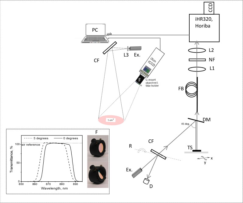
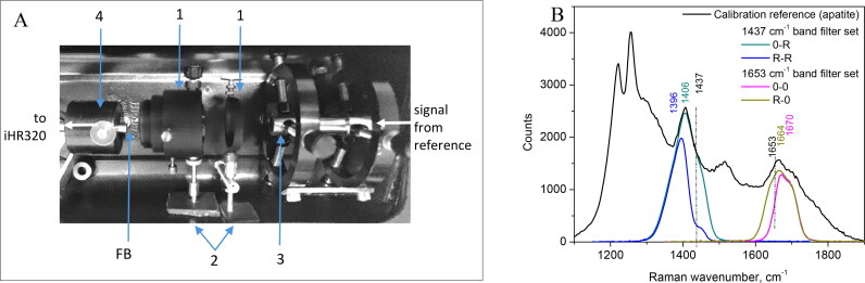
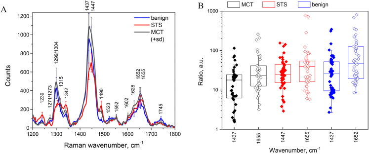
This study introduces Raman imaging technique for diagnosing skin cancer in veterinary oncology patients (dogs and cats). Initially, Raman spectral bands (with specificity to certain molecular structures and functional groups) were identified in formalin-fixed samples of mast cell tumors and soft tissue sarcomas, obtained through routine veterinary biopsy submissions. Then, a custom-built Raman macro-imaging system featuring an intensified CCD camera (iXon Ultra 888, Andor, UK), tunable narrow-band Semrock (USA) optical filter compartment was used to map the spectral features at 1437 cm-1 and 1655 cm-1 in ex vivo tissue. This approach enabled wide-field (cm2), rapid (within seconds), and safe (< 400 mW/cm2) imaging conditions, supporting accurate diagnosis of tissue state. The findings indicate that machine learning classifiers - particularly support vector machine (SVM) and decision tree (DT) - effectively distinguished between soft tissue sarcoma, mastocytoma and benign tissues using Raman spectral band imaging data. Additionally, combining Raman macro-imaging with residual near-infrared (NIR) autofluorescence as a bimodal imaging technique enhanced diagnostic performance, reaching 85 - 95% in accuracy, sensitivity, specificity, and precision - even with a single spectral band (1437 cm-1 or 1655 cm-1). In conclusion, the proposed bi-modal imaging is a pioneering method for veterinary oncology science, offering to improve the diagnostic accuracy of malignant tumors.
期刊介绍:
Veterinary Quarterly is an international open access journal which publishes high quality review articles and original research in the field of veterinary science and animal diseases. The journal publishes research on a range of different animal species and topics including: - Economically important species such as domesticated and non-domesticated farm animals, including avian and poultry diseases; - Companion animals (dogs, cats, horses, pocket pets and exotics); - Wildlife species; - Infectious diseases; - Diagnosis; - Treatment including pharmacology and vaccination
 求助内容:
求助内容: 应助结果提醒方式:


