{"title":"Exploring the Effects of Action Observation Therapy on Swallowing Disorders in Stroke: A Functional Connectivity-Based fMRI Study.","authors":"Xuting Chen, Xiaolin Sun, Fang Shen, Zhongli Wang, Meihong Zhu, Jianming Fu, Yunhai Yao, Jie Wang, Linhua Tao, Lianjie Ma, Ming Zeng, Xudong Gu","doi":"10.1155/np/8176431","DOIUrl":null,"url":null,"abstract":"<p><p><b>Objective:</b> This study aims to investigate the impact of action observation therapy (AOT) on swallowing disorders following a stroke. Utilizing functional magnetic resonance imaging (fMRI) technology, the study will examine adjustments in brain activity and functional connectivity (FC), providing novel insights for the rehabilitation of swallowing function in stroke patients. <b>Methods:</b> In this study, 11 healthy controls (HCs) and 11 stroke patients were included. The stroke patients underwent a 4-week AOT. To assess the differences in brain region activity between the patients before and after treatment and the HCs, regional homogeneity (ReHo), and degree centrality (DC) were calculated based on fMRI data separately. Important brain regions were selected as regions of interest (ROIs) for subsequent FC analysis, and finally, comparisons were made to evaluate the therapeutic effects. <b>Results:</b> Comparing stroke patients before treatment with HCs, the ReHo values were relatively higher in the inferior temporal gyrus, median cingulate, and paracingulate gyri, and relatively lower in the calcarine fissure and surrounding cortex, middle occipital gyrus, and paracentral lobule. The DC values were relatively higher in the cerebellum, middle frontal gyrus, inferior temporal gyrus, inferior frontal gyrus, orbital part, middle frontal gyrus, and supramarginal gyrus, and relatively lower in the cuneus and paracentral lobule. The FC between the parahippocampal gyrus and the superior parietal gyrus was relatively high, and the FC between the superior occipital gyrus and the superior parietal gyrus was relatively low. Comparing stroke patients after treatment with HCs, the ReHo values were relatively higher in the caudate nucleus, and relatively lower in the cerebellum, superior frontal gyrus, medial orbital, calcarine fissure and surrounding cortex, and middle temporal gyrus. The DC values were relatively higher in the middle frontal gyrus and superior frontal gyrus, and relatively lower in the temporal pole: superior temporal gyrus, calcarine fissure, and surrounding cortex. The FC between the caudate nucleus and the superior parietal gyrus was relatively high, and the FC between the calcarine fissure and surrounding cortex, middle frontal gyrus, orbital part, and the superior parietal gyrus was relatively low. There was no significant difference in ReHo values between stroke patients before and after treatment. The DC value in the superior parietal gyrus increased, and the FC in the superior parietal gyrus and precuneus gyrus was also significantly enhanced before and after treatment. <b>Conclusion:</b> The results of this study indicate that the AOT has a positive effect on enhancing the functional connection and information transmission capabilities of specific brain regions. The impact of this therapy on brain function helps us understand the potential mechanisms of swallowing function network reorganization deeper. <b>Trial Registration:</b> Chinese Clinical Trial Registry: ChiCTR1900021849.</p>","PeriodicalId":19122,"journal":{"name":"Neural Plasticity","volume":"2025 ","pages":"8176431"},"PeriodicalIF":3.7000,"publicationDate":"2025-03-31","publicationTypes":"Journal Article","fieldsOfStudy":null,"isOpenAccess":false,"openAccessPdf":"https://www.ncbi.nlm.nih.gov/pmc/articles/PMC11976039/pdf/","citationCount":"0","resultStr":null,"platform":"Semanticscholar","paperid":null,"PeriodicalName":"Neural Plasticity","FirstCategoryId":"3","ListUrlMain":"https://doi.org/10.1155/np/8176431","RegionNum":4,"RegionCategory":"医学","ArticlePicture":[],"TitleCN":null,"AbstractTextCN":null,"PMCID":null,"EPubDate":"2025/1/1 0:00:00","PubModel":"eCollection","JCR":"Q2","JCRName":"Medicine","Score":null,"Total":0}
引用次数: 0
Abstract
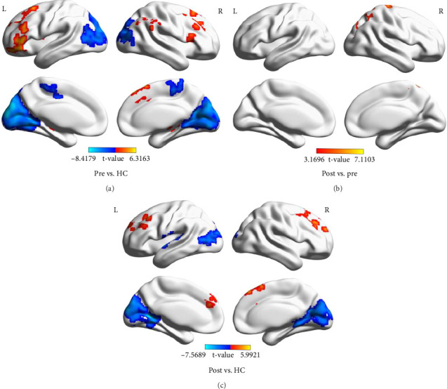
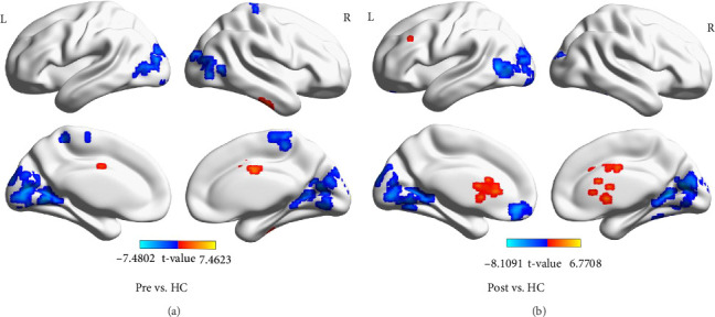
Objective: This study aims to investigate the impact of action observation therapy (AOT) on swallowing disorders following a stroke. Utilizing functional magnetic resonance imaging (fMRI) technology, the study will examine adjustments in brain activity and functional connectivity (FC), providing novel insights for the rehabilitation of swallowing function in stroke patients. Methods: In this study, 11 healthy controls (HCs) and 11 stroke patients were included. The stroke patients underwent a 4-week AOT. To assess the differences in brain region activity between the patients before and after treatment and the HCs, regional homogeneity (ReHo), and degree centrality (DC) were calculated based on fMRI data separately. Important brain regions were selected as regions of interest (ROIs) for subsequent FC analysis, and finally, comparisons were made to evaluate the therapeutic effects. Results: Comparing stroke patients before treatment with HCs, the ReHo values were relatively higher in the inferior temporal gyrus, median cingulate, and paracingulate gyri, and relatively lower in the calcarine fissure and surrounding cortex, middle occipital gyrus, and paracentral lobule. The DC values were relatively higher in the cerebellum, middle frontal gyrus, inferior temporal gyrus, inferior frontal gyrus, orbital part, middle frontal gyrus, and supramarginal gyrus, and relatively lower in the cuneus and paracentral lobule. The FC between the parahippocampal gyrus and the superior parietal gyrus was relatively high, and the FC between the superior occipital gyrus and the superior parietal gyrus was relatively low. Comparing stroke patients after treatment with HCs, the ReHo values were relatively higher in the caudate nucleus, and relatively lower in the cerebellum, superior frontal gyrus, medial orbital, calcarine fissure and surrounding cortex, and middle temporal gyrus. The DC values were relatively higher in the middle frontal gyrus and superior frontal gyrus, and relatively lower in the temporal pole: superior temporal gyrus, calcarine fissure, and surrounding cortex. The FC between the caudate nucleus and the superior parietal gyrus was relatively high, and the FC between the calcarine fissure and surrounding cortex, middle frontal gyrus, orbital part, and the superior parietal gyrus was relatively low. There was no significant difference in ReHo values between stroke patients before and after treatment. The DC value in the superior parietal gyrus increased, and the FC in the superior parietal gyrus and precuneus gyrus was also significantly enhanced before and after treatment. Conclusion: The results of this study indicate that the AOT has a positive effect on enhancing the functional connection and information transmission capabilities of specific brain regions. The impact of this therapy on brain function helps us understand the potential mechanisms of swallowing function network reorganization deeper. Trial Registration: Chinese Clinical Trial Registry: ChiCTR1900021849.
期刊介绍:
Neural Plasticity is an international, interdisciplinary journal dedicated to the publication of articles related to all aspects of neural plasticity, with special emphasis on its functional significance as reflected in behavior and in psychopathology. Neural Plasticity publishes research and review articles from the entire range of relevant disciplines, including basic neuroscience, behavioral neuroscience, cognitive neuroscience, biological psychology, and biological psychiatry.
 求助内容:
求助内容: 应助结果提醒方式:


