Comparison of TLR4, NF-κB and IRF3 expression in kidney tissue between lupus nephritis (LN) and systemic lupus erythematosus (SLE): a pristane-induced lupus mice model study.
Yuswanto Setyawan, Hani Susianti, Nur Samsu, Loeki Enggar Fitri
{"title":"Comparison of TLR4, NF-κB and IRF3 expression in kidney tissue between lupus nephritis (LN) and systemic lupus erythematosus (SLE): a pristane-induced lupus mice model study.","authors":"Yuswanto Setyawan, Hani Susianti, Nur Samsu, Loeki Enggar Fitri","doi":"10.1136/lupus-2024-001445","DOIUrl":null,"url":null,"abstract":"<p><strong>Introduction and purpose: </strong>Lupus nephritis (LN) is a major cause of morbidity and mortality in patients with SLE, a complex autoimmune disease characterised by loss of tolerance to self-nuclear antigens. Toll-like receptor 4 (TLR4), the first line of defence in the innate immune system, has been linked to the pathogenesis of autoimmune diseases and LN by activating nuclear factor-κB (NF-κB) or interferon regulatory transcription factor 3 (IRF3). Local expression of those biomarkers in pristane-induced lupus mice is still unknown. Therefore, this study aimed to prove the role of TLR4, NF-κB and IRF3 in pristane-induced lupus mice.</p><p><strong>Subjects and methods: </strong>The study subjects were female Balb/c pristane-induced lupus mice model, 8-12 weeks of age, n=30, divided into two groups, nephritis (LN group) and non-nephritis (SLE group). The control group were age-matched healthy female Balb/c mice, n=11. All mice were euthanised at weeks 16. Kidney tissue was taken for histopathology examination and TLR4, NF-κB, IRF3 immunofluorescence assay. The diagnosis of LN was based on proteinuria and histopathology examination according to the ISN/RPS 2004 classification of LN. Statistical analysis was performed using IBM SPSS Statistics V.25. P value <0.05 was considered statistically significant.</p><p><strong>Results: </strong>There were significant differences in the expressions of TLR4, NF-κB and IRF3 among the LN, SLE and healthy control groups (p=0.000), with the highest expression found in the LN group for all markers. The linear regression between TLR4 and NF-κB resulted in p value=0.000; R<sup>2</sup>=0.817; β=0.904. Linear regression between TLR4 and IRF3 showed p value=0.000; R<sup>2</sup>=0.896; β=0.947, which means TLR4 had an 81.7% effect on NF-κB and 89.6% on IRF3 expression.</p><p><strong>Conclusion: </strong>TLR4, NF-κB and IRF3 expression were increased in lupus, with the highest expression found in the LN group, suggesting that these biomarkers may be responsible for the development of nephritis in SLE, with TLR4 likely playing a dominant role in this pathway. Increased expression of these biomarkers in lupus without nephritis may indicate progression towards nephritis, which still needs to be proven with further research.</p>","PeriodicalId":18126,"journal":{"name":"Lupus Science & Medicine","volume":"12 1","pages":""},"PeriodicalIF":3.5000,"publicationDate":"2025-04-07","publicationTypes":"Journal Article","fieldsOfStudy":null,"isOpenAccess":false,"openAccessPdf":"https://www.ncbi.nlm.nih.gov/pmc/articles/PMC11977469/pdf/","citationCount":"0","resultStr":null,"platform":"Semanticscholar","paperid":null,"PeriodicalName":"Lupus Science & Medicine","FirstCategoryId":"3","ListUrlMain":"https://doi.org/10.1136/lupus-2024-001445","RegionNum":2,"RegionCategory":"医学","ArticlePicture":[],"TitleCN":null,"AbstractTextCN":null,"PMCID":null,"EPubDate":"","PubModel":"","JCR":"Q1","JCRName":"RHEUMATOLOGY","Score":null,"Total":0}
引用次数: 0
Abstract
Introduction and purpose: Lupus nephritis (LN) is a major cause of morbidity and mortality in patients with SLE, a complex autoimmune disease characterised by loss of tolerance to self-nuclear antigens. Toll-like receptor 4 (TLR4), the first line of defence in the innate immune system, has been linked to the pathogenesis of autoimmune diseases and LN by activating nuclear factor-κB (NF-κB) or interferon regulatory transcription factor 3 (IRF3). Local expression of those biomarkers in pristane-induced lupus mice is still unknown. Therefore, this study aimed to prove the role of TLR4, NF-κB and IRF3 in pristane-induced lupus mice.
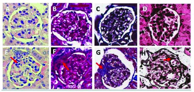
Subjects and methods: The study subjects were female Balb/c pristane-induced lupus mice model, 8-12 weeks of age, n=30, divided into two groups, nephritis (LN group) and non-nephritis (SLE group). The control group were age-matched healthy female Balb/c mice, n=11. All mice were euthanised at weeks 16. Kidney tissue was taken for histopathology examination and TLR4, NF-κB, IRF3 immunofluorescence assay. The diagnosis of LN was based on proteinuria and histopathology examination according to the ISN/RPS 2004 classification of LN. Statistical analysis was performed using IBM SPSS Statistics V.25. P value <0.05 was considered statistically significant.
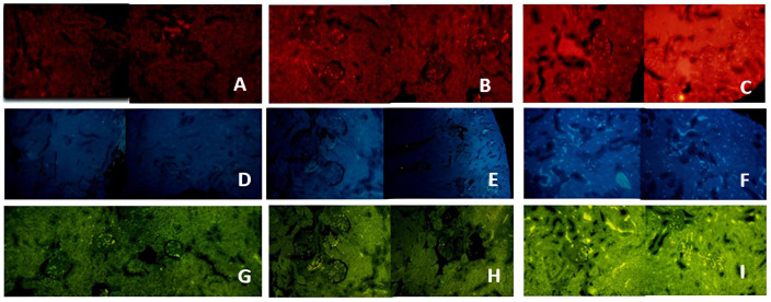
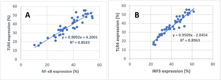
Results: There were significant differences in the expressions of TLR4, NF-κB and IRF3 among the LN, SLE and healthy control groups (p=0.000), with the highest expression found in the LN group for all markers. The linear regression between TLR4 and NF-κB resulted in p value=0.000; R2=0.817; β=0.904. Linear regression between TLR4 and IRF3 showed p value=0.000; R2=0.896; β=0.947, which means TLR4 had an 81.7% effect on NF-κB and 89.6% on IRF3 expression.
Conclusion: TLR4, NF-κB and IRF3 expression were increased in lupus, with the highest expression found in the LN group, suggesting that these biomarkers may be responsible for the development of nephritis in SLE, with TLR4 likely playing a dominant role in this pathway. Increased expression of these biomarkers in lupus without nephritis may indicate progression towards nephritis, which still needs to be proven with further research.
期刊介绍:
Lupus Science & Medicine is a global, peer reviewed, open access online journal that provides a central point for publication of basic, clinical, translational, and epidemiological studies of all aspects of lupus and related diseases. It is the first lupus-specific open access journal in the world and was developed in response to the need for a barrier-free forum for publication of groundbreaking studies in lupus. The journal publishes research on lupus from fields including, but not limited to: rheumatology, dermatology, nephrology, immunology, pediatrics, cardiology, hepatology, pulmonology, obstetrics and gynecology, and psychiatry.
 求助内容:
求助内容: 应助结果提醒方式:


