Edgar Buloz-Osorio, Marisa Ortega-Sánchez, Miguel B Royo-Salvador, Alfonso Rodríguez-Baeza
{"title":"Morphological analysis of the filum terminale and detailed description of the distal filum terminale externum: a cadaveric study.","authors":"Edgar Buloz-Osorio, Marisa Ortega-Sánchez, Miguel B Royo-Salvador, Alfonso Rodríguez-Baeza","doi":"10.3389/fnana.2025.1547165","DOIUrl":null,"url":null,"abstract":"<p><p>This observational, descriptive anatomical cadaveric study aimed to identify, characterize, and analyze the morphometric parameters of the filum terminale (FT) and macroscopically describe the distal insertion of the FTE. The FT is a complex, three-dimensional, fibro-cellular structure of connective tissue and glial cells, extending from the conus medullaris (CM) to the dural sac (DS) and coccyx. It is divided into two segments: an intradural filum terminale internum (FTI) and extradural filum terminale externum (FTE). Few studies have comprehensively addressed its morphometric characteristics in the last decades. Thirty-eight embalmed (M = 16, F = 22) human cadavers were examined to determine the CM-FTI and DS-FTE vertebral levels and FT, FTI, and FTE lengths and widths. FTI and FTE segmental diameters, correlations, gross characteristics, tension, and mobility <i>in situ</i> and <i>ex vivo</i> were assessed. FTE distal insertion is thoroughly described. FT, FTI, and FTE mean lengths were 254.32 mm (±26.46), 152.75 mm (±22.02), and 106.64 mm (±12.21), respectively. The CM-FTI junction was observed at the L1-L2 disk space (32.1%), DS-FTE fusion in the upper third of S2 (39.3%), and FTI-DS fusion in the DS midline (46.4%). FT length and FTI, FTE lengths were directly correlated, as were all FTI diameters. FT gross characteristics were an irregular surface (71.4%), bright hue (57.1%), macroscopic FTI-CM contrast (64.3%), filiform shape (60.7%), and movement-resistance (53.6%). The FTE exhibited a flattened shape (64.3%), immobility (60.7%), distal insertion at Cx1 (67.8%) and one distal strand (60.7%). FTI segments ≥ 2 mm were uncommon (21.4%). The FTE distal insertion is variable, inserting as strands, with vascular tissue surrounding it. A distal coccygeal venous plexus and single or multiple strand-like insertions of the distal FTE are for the first time described in detail. Discrepancies in the morphometric parameters of the FT between studies highlight the need for standardized protocols, especially given the FT's anatomic-clinical importance and potential role as a neural progenitor niche. We provide a comprehensive basis for future standardized morphometric analyses, acknowledging the limitations of embalmed cadaveric studies.</p>","PeriodicalId":12572,"journal":{"name":"Frontiers in Neuroanatomy","volume":"19 ","pages":"1547165"},"PeriodicalIF":2.3000,"publicationDate":"2025-03-25","publicationTypes":"Journal Article","fieldsOfStudy":null,"isOpenAccess":false,"openAccessPdf":"https://www.ncbi.nlm.nih.gov/pmc/articles/PMC11975916/pdf/","citationCount":"0","resultStr":null,"platform":"Semanticscholar","paperid":null,"PeriodicalName":"Frontiers in Neuroanatomy","FirstCategoryId":"3","ListUrlMain":"https://doi.org/10.3389/fnana.2025.1547165","RegionNum":4,"RegionCategory":"医学","ArticlePicture":[],"TitleCN":null,"AbstractTextCN":null,"PMCID":null,"EPubDate":"2025/1/1 0:00:00","PubModel":"eCollection","JCR":"Q1","JCRName":"ANATOMY & MORPHOLOGY","Score":null,"Total":0}
引用次数: 0
Abstract
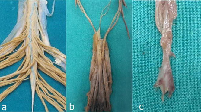
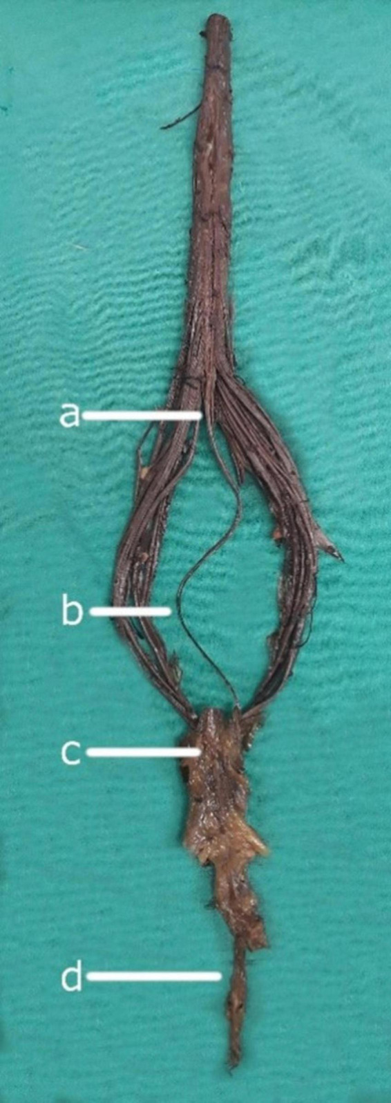
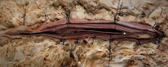
This observational, descriptive anatomical cadaveric study aimed to identify, characterize, and analyze the morphometric parameters of the filum terminale (FT) and macroscopically describe the distal insertion of the FTE. The FT is a complex, three-dimensional, fibro-cellular structure of connective tissue and glial cells, extending from the conus medullaris (CM) to the dural sac (DS) and coccyx. It is divided into two segments: an intradural filum terminale internum (FTI) and extradural filum terminale externum (FTE). Few studies have comprehensively addressed its morphometric characteristics in the last decades. Thirty-eight embalmed (M = 16, F = 22) human cadavers were examined to determine the CM-FTI and DS-FTE vertebral levels and FT, FTI, and FTE lengths and widths. FTI and FTE segmental diameters, correlations, gross characteristics, tension, and mobility in situ and ex vivo were assessed. FTE distal insertion is thoroughly described. FT, FTI, and FTE mean lengths were 254.32 mm (±26.46), 152.75 mm (±22.02), and 106.64 mm (±12.21), respectively. The CM-FTI junction was observed at the L1-L2 disk space (32.1%), DS-FTE fusion in the upper third of S2 (39.3%), and FTI-DS fusion in the DS midline (46.4%). FT length and FTI, FTE lengths were directly correlated, as were all FTI diameters. FT gross characteristics were an irregular surface (71.4%), bright hue (57.1%), macroscopic FTI-CM contrast (64.3%), filiform shape (60.7%), and movement-resistance (53.6%). The FTE exhibited a flattened shape (64.3%), immobility (60.7%), distal insertion at Cx1 (67.8%) and one distal strand (60.7%). FTI segments ≥ 2 mm were uncommon (21.4%). The FTE distal insertion is variable, inserting as strands, with vascular tissue surrounding it. A distal coccygeal venous plexus and single or multiple strand-like insertions of the distal FTE are for the first time described in detail. Discrepancies in the morphometric parameters of the FT between studies highlight the need for standardized protocols, especially given the FT's anatomic-clinical importance and potential role as a neural progenitor niche. We provide a comprehensive basis for future standardized morphometric analyses, acknowledging the limitations of embalmed cadaveric studies.
期刊介绍:
Frontiers in Neuroanatomy publishes rigorously peer-reviewed research revealing important aspects of the anatomical organization of all nervous systems across all species. Specialty Chief Editor Javier DeFelipe at the Cajal Institute (CSIC) is supported by an outstanding Editorial Board of international experts. This multidisciplinary open-access journal is at the forefront of disseminating and communicating scientific knowledge and impactful discoveries to researchers, academics, clinicians and the public worldwide.
 求助内容:
求助内容: 应助结果提醒方式:


