{"title":"Evaluation of CE-MATRIX-Enhanced FLAIR imaging in the detection of leptomeningeal metastasis.","authors":"Junhui Yuan, Shaobo Fang, Fan Meng, Yue Wu, Dongqiu Shan, Chunmiao Xu, Renzhi Zhang, Xuejun Chen","doi":"10.1186/s40644-025-00867-z","DOIUrl":null,"url":null,"abstract":"<p><strong>Objective: </strong>To investigate the diagnostic value of CE-MATRIX-T1FLAIR and 3D CE-T2FLAIR sequences based on Contrast Enhancement Modulated flip Angle Technique in Refocused Imaging with eXtended echo train (CE-MATRIX) technology for detecting Leptomeningeal Metastasis (LM) using Fluid Attenuated Inversion Recovery (FLAIR) imaging.</p><p><strong>Methods: </strong>This prospective study included 563 hospitalized patients with clinically suspected LM, diagnosed with malignant tumors between January 2022 and October 2023 at Henan Cancer Hospital. Both CE-MATRIX-T1FLAIR and 3D CE-T2FLAIR sequences were used for imaging. Two radiologists independently evaluated image quality, diagnostic confidence, and objective measurements, diagnosing LM as positive or negative, with disagreements resolved by consultation. Subjective and objective scores were compared using the Wilcoxon signed-rank test. The diagnostic performance of the sequences was compared using ROC curve analysis, with cerebrospinal fluid (CSF) cytology as the gold standard. Sensitivity, specificity, positive predictive value (PPV), negative predictive value (NPV), accuracy, and area under the curve (AUC) values were calculated and compared using Z-tests.</p><p><strong>Results: </strong>LM was confirmed in 321 patients. CE-MATRIX-T1FLAIR showed superior subjective scores in image quality and diagnostic confidence (p < 0.001). Though CE-MATRIX-T1FLAIR had a lower SNR (p = 0.013), it demonstrated higher sensitivity, specificity, PPV, NPV, accuracy, and AUC than 3D CE-T2FLAIR (p < 0.001). Both sequences provided effective diagnosis and differentiation of LM.</p><p><strong>Conclusion: </strong>CE-MATRIX-T1FLAIR offers superior diagnostic performance compared to 3D CE-T2FLAIR for LM, with slightly better subjective ratings despite a lower SNR. Both sequences are effective for diagnosing LM.</p>","PeriodicalId":9548,"journal":{"name":"Cancer Imaging","volume":"25 1","pages":"53"},"PeriodicalIF":3.5000,"publicationDate":"2025-04-08","publicationTypes":"Journal Article","fieldsOfStudy":null,"isOpenAccess":false,"openAccessPdf":"https://www.ncbi.nlm.nih.gov/pmc/articles/PMC11980099/pdf/","citationCount":"0","resultStr":null,"platform":"Semanticscholar","paperid":null,"PeriodicalName":"Cancer Imaging","FirstCategoryId":"3","ListUrlMain":"https://doi.org/10.1186/s40644-025-00867-z","RegionNum":2,"RegionCategory":"医学","ArticlePicture":[],"TitleCN":null,"AbstractTextCN":null,"PMCID":null,"EPubDate":"","PubModel":"","JCR":"Q2","JCRName":"ONCOLOGY","Score":null,"Total":0}
引用次数: 0
Abstract
Objective: To investigate the diagnostic value of CE-MATRIX-T1FLAIR and 3D CE-T2FLAIR sequences based on Contrast Enhancement Modulated flip Angle Technique in Refocused Imaging with eXtended echo train (CE-MATRIX) technology for detecting Leptomeningeal Metastasis (LM) using Fluid Attenuated Inversion Recovery (FLAIR) imaging.
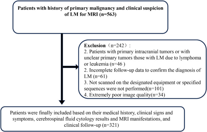
Methods: This prospective study included 563 hospitalized patients with clinically suspected LM, diagnosed with malignant tumors between January 2022 and October 2023 at Henan Cancer Hospital. Both CE-MATRIX-T1FLAIR and 3D CE-T2FLAIR sequences were used for imaging. Two radiologists independently evaluated image quality, diagnostic confidence, and objective measurements, diagnosing LM as positive or negative, with disagreements resolved by consultation. Subjective and objective scores were compared using the Wilcoxon signed-rank test. The diagnostic performance of the sequences was compared using ROC curve analysis, with cerebrospinal fluid (CSF) cytology as the gold standard. Sensitivity, specificity, positive predictive value (PPV), negative predictive value (NPV), accuracy, and area under the curve (AUC) values were calculated and compared using Z-tests.
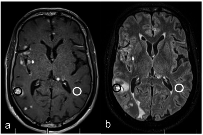
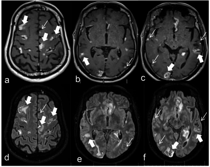
Results: LM was confirmed in 321 patients. CE-MATRIX-T1FLAIR showed superior subjective scores in image quality and diagnostic confidence (p < 0.001). Though CE-MATRIX-T1FLAIR had a lower SNR (p = 0.013), it demonstrated higher sensitivity, specificity, PPV, NPV, accuracy, and AUC than 3D CE-T2FLAIR (p < 0.001). Both sequences provided effective diagnosis and differentiation of LM.
Conclusion: CE-MATRIX-T1FLAIR offers superior diagnostic performance compared to 3D CE-T2FLAIR for LM, with slightly better subjective ratings despite a lower SNR. Both sequences are effective for diagnosing LM.
Cancer ImagingONCOLOGY-RADIOLOGY, NUCLEAR MEDICINE & MEDICAL IMAGING
CiteScore
7.00
自引率
0.00%
发文量
66
审稿时长
>12 weeks
期刊介绍:
Cancer Imaging is an open access, peer-reviewed journal publishing original articles, reviews and editorials written by expert international radiologists working in oncology.
The journal encompasses CT, MR, PET, ultrasound, radionuclide and multimodal imaging in all kinds of malignant tumours, plus new developments, techniques and innovations. Topics of interest include:
Breast Imaging
Chest
Complications of treatment
Ear, Nose & Throat
Gastrointestinal
Hepatobiliary & Pancreatic
Imaging biomarkers
Interventional
Lymphoma
Measurement of tumour response
Molecular functional imaging
Musculoskeletal
Neuro oncology
Nuclear Medicine
Paediatric.

 求助内容:
求助内容: 应助结果提醒方式:


