Ion-conducting and gating molecular mechanisms of channelrhodopsin revealed by true-atomic-resolution structures of open and closed states
IF 10.1
1区 生物学
Q1 BIOCHEMISTRY & MOLECULAR BIOLOGY
引用次数: 0
Abstract
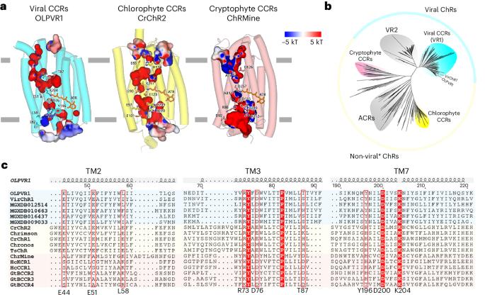
Channelrhodopsins (ChRs) have emerged as major optogenetics tools, particularly in neuroscience. Despite their importance, the molecular mechanism of ChR opening remains elusive. Moreover, all reported structures of ChRs correspond to either a closed or an early intermediate state and lack the necessary level of detail owing to the limited resolution. Here we present the structures of the closed and open states of a cation-conducting ChR, OLPVR1, from Organic Lake phycodnavirus, belonging to the family of viral ChRs solved at 1.1- and 1.3-Å resolution at physiologically relevant pH conditions (pH 8.0). OLPVR1 was expressed in Escherichia coli and crystallized using an in meso approach, and the structures were solved by X-ray crystallography. We also present the structure of the OLPVR1 protonated state at acidic pH (pH 2.5) at 1.4-Å resolution. Together, these three structures elucidate the molecular mechanisms of the channel’s opening and permeability in detail. Extensive functional studies support the proposed mechanisms. Channel opening is controlled by isomerization of the retinal cofactor, triggering protonation of proton acceptors and deprotonation of proton donors located in the three gates of the channel. The E51 residue in the core of the central gate (similar to E90 of ChR2 from Chlamydomonas reinhardtii) plays a key role in the opening of the channel. E51 flips out of the gate and towards the proton acceptor D200 (D253 in ChR2 in C. reinhardtii), establishing a hydrogen bond between them. Despite differences in subfamilies of ChRs, they share a common gate–cavity architecture, suggesting that they could have similar general gating mechanisms. These results enabled us to design viral rhodopsin with improved properties for optogenetic applications. The structural data and mechanisms might also be helpful for better understanding other ChRs and their engineering. Channelrhodopsins (ChRs) are primary tools for precise optical control over living cells. Structures of a viral ChR, OLPVR1, in closed (1.1 Å) and open (1.3 Å) states reveal the key details of its molecular mechanism.

通道紫红质的离子传导和门控分子机制揭示的真原子分辨率结构的开放和关闭状态
通道视紫红质(ChRs)已成为主要的光遗传学工具,特别是在神经科学领域。尽管它们很重要,但ChR打开的分子机制仍然难以捉摸。此外,所有报道的chr结构都对应于封闭或早期中间状态,由于分辨率有限,缺乏必要的细节水平。本文研究了有机湖藻病毒的阳离子导电ChR OLPVR1的封闭和开放状态结构,该病毒属于在生理相关的pH条件下(pH 8.0)以1.1-和1.3-Å分辨率溶解的病毒ChR家族。OLPVR1在大肠杆菌中表达,用中介观法结晶,并用x射线晶体学解析其结构。我们还以1.4-Å分辨率展示了酸性pH (pH 2.5)下OLPVR1质子化状态的结构。总之,这三种结构详细阐明了通道打开和渗透的分子机制。广泛的功能研究支持所提出的机制。通道打开由视网膜辅助因子的异构化控制,触发位于通道三个门的质子受体的质子化和质子供体的去质子化。中央通道核心的E51残基(类似莱茵衣藻ChR2的E90残基)在通道打开中起关键作用。E51跃出大门,奔向质子受体D200 (C. reinhardtii中ChR2中的D253),在它们之间建立了氢键。尽管chr亚家族存在差异,但它们具有共同的门腔结构,这表明它们可能具有相似的一般门机制。这些结果使我们能够设计具有改进性质的病毒视紫红质用于光遗传学应用。结构数据和机制也可能有助于更好地了解其他chr及其工程。
本文章由计算机程序翻译,如有差异,请以英文原文为准。
求助全文
约1分钟内获得全文
求助全文
来源期刊
Nature Structural & Molecular Biology
BIOCHEMISTRY & MOLECULAR BIOLOGY-BIOPHYSICS
CiteScore
22.00
自引率
1.80%
发文量
160
审稿时长
3-8 weeks
期刊介绍:
Nature Structural & Molecular Biology is a comprehensive platform that combines structural and molecular research. Our journal focuses on exploring the functional and mechanistic aspects of biological processes, emphasizing how molecular components collaborate to achieve a particular function. While structural data can shed light on these insights, our publication does not require them as a prerequisite.
 求助内容:
求助内容: 应助结果提醒方式:


