Fernando César Moreira, Helder Baldi Jacob, Guilherme Dos Reis Pereira Janson, Daniela Gamba Garib
{"title":"Predicted and final tooth position assessment following indirect bonding planned by a digital system.","authors":"Fernando César Moreira, Helder Baldi Jacob, Guilherme Dos Reis Pereira Janson, Daniela Gamba Garib","doi":"10.1590/2177-6709.30.1.e252451.oar","DOIUrl":null,"url":null,"abstract":"<p><strong>Introduction: </strong>The purpose of this study was to evaluate the agreement between the predicted and the achieved tooth position planned by an orthodontic digital system.</p><p><strong>Methods: </strong>Digital models of the setup (Predicted) and the treated (Treated) groups of 23 subjects with Class I malocclusion were obtained. Digital models (Predicted and Treated) of each patient were superimposed, and referential geometric planes were constructed for linear and angular measurements: arch perimeter, arch depth, intercanine and intermolar widths, mesiodistal crown angulation, and buccolingual crown inclination. Bland-Altman analysis was performed to establish the agreement between the measurements. Spearman's correlation coefficient was used to evaluate the correlation between groups.</p><p><strong>Results: </strong>Compared to Predicted group, the Treated group presented larger linear measurements for all measurements: 1) arch perimeter: 1.77±2.10 mm (maxilla) and 1.78±1.74 mm (mandible); 2) arch depth: 0.50±0.69 mm (maxilla) and 0.38±0.81 mm (mandible); 3) intercanine width: 0.30±0.98 mm (maxilla) and 0.49±0.64 mm (mandible), and; 4) intermolar width: 0.70±1.63 mm (maxilla) and 1.13±1.62 mm (mandible). Seven out of 14 angular measurements showed statistical differences between Predicted and Treated groups in the maxilla, while six out of 14 angular measurements were statistically significant between the two groups; the differences ranging from -8.91º to 1.91º and from -3.53° to 9.59° in the maxilla and mandible, respectively.</p><p><strong>Conclusions: </strong>The agreement between the Predicted and Treated groups was majority within the limits. The predictions of the digital system were not accurate in some parameters; however, most of the differences were within clinical acceptable range. Although there are some inaccuracies, the limitations do not seem to interfere with clinical outcomes and the quality of the treatment.</p>","PeriodicalId":38720,"journal":{"name":"Dental Press Journal of Orthodontics","volume":"30 1","pages":"e252451"},"PeriodicalIF":0.0000,"publicationDate":"2025-04-07","publicationTypes":"Journal Article","fieldsOfStudy":null,"isOpenAccess":false,"openAccessPdf":"https://www.ncbi.nlm.nih.gov/pmc/articles/PMC11980639/pdf/","citationCount":"0","resultStr":null,"platform":"Semanticscholar","paperid":null,"PeriodicalName":"Dental Press Journal of Orthodontics","FirstCategoryId":"1085","ListUrlMain":"https://doi.org/10.1590/2177-6709.30.1.e252451.oar","RegionNum":0,"RegionCategory":null,"ArticlePicture":[],"TitleCN":null,"AbstractTextCN":null,"PMCID":null,"EPubDate":"2025/1/1 0:00:00","PubModel":"eCollection","JCR":"Q2","JCRName":"Medicine","Score":null,"Total":0}
引用次数: 0
Abstract
Introduction: The purpose of this study was to evaluate the agreement between the predicted and the achieved tooth position planned by an orthodontic digital system.
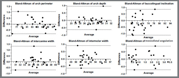
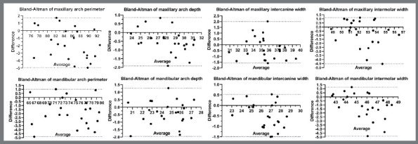
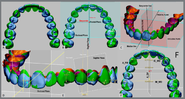
Methods: Digital models of the setup (Predicted) and the treated (Treated) groups of 23 subjects with Class I malocclusion were obtained. Digital models (Predicted and Treated) of each patient were superimposed, and referential geometric planes were constructed for linear and angular measurements: arch perimeter, arch depth, intercanine and intermolar widths, mesiodistal crown angulation, and buccolingual crown inclination. Bland-Altman analysis was performed to establish the agreement between the measurements. Spearman's correlation coefficient was used to evaluate the correlation between groups.
Results: Compared to Predicted group, the Treated group presented larger linear measurements for all measurements: 1) arch perimeter: 1.77±2.10 mm (maxilla) and 1.78±1.74 mm (mandible); 2) arch depth: 0.50±0.69 mm (maxilla) and 0.38±0.81 mm (mandible); 3) intercanine width: 0.30±0.98 mm (maxilla) and 0.49±0.64 mm (mandible), and; 4) intermolar width: 0.70±1.63 mm (maxilla) and 1.13±1.62 mm (mandible). Seven out of 14 angular measurements showed statistical differences between Predicted and Treated groups in the maxilla, while six out of 14 angular measurements were statistically significant between the two groups; the differences ranging from -8.91º to 1.91º and from -3.53° to 9.59° in the maxilla and mandible, respectively.
Conclusions: The agreement between the Predicted and Treated groups was majority within the limits. The predictions of the digital system were not accurate in some parameters; however, most of the differences were within clinical acceptable range. Although there are some inaccuracies, the limitations do not seem to interfere with clinical outcomes and the quality of the treatment.
期刊介绍:
The Dental Press Journal of Orthodontics publishes scientific research articles, significant reviews, clinical and technical case reports, brief communications, and other materials related to Orthodontics and Facial Orthopedics.

 求助内容:
求助内容: 应助结果提醒方式:


