Masayuki Shintaku, Tokiko Okunobo, Hiroki Nakamura, Takashi Doi, Akira Tanaka, Koji Tsuta
{"title":"Primary Eosinophilic Panniculitis of the Greater Omentum in a Young Girl: A Case Report.","authors":"Masayuki Shintaku, Tokiko Okunobo, Hiroki Nakamura, Takashi Doi, Akira Tanaka, Koji Tsuta","doi":"10.1159/000544861","DOIUrl":null,"url":null,"abstract":"<p><strong>Introduction: </strong>Primary (or idiopathic) panniculitis involving the intra-abdominal adipose tissue is rare, and its pathogenesis remains unknown. A case of primary eosinophilic panniculitis that involved the greater omentum of a girl is reported.</p><p><strong>Case presentation: </strong>The patient, an 11-year-old girl, complained of dull periumbilical pain and nausea, and radiological examination showed a mass lesion in the abdomino-pelvic cavity. On laparoscopy, a plaque-like, flat mass was seen in the greater omentum, and laparoscopic omental resection was performed. On histopathological examination, the interlobular fibrous septa of omental adipose tissue were widened by inflammatory edema, prominent infiltration of eosinophils, and loose proliferation of myofibroblasts. Dense lymphocytic infiltration was also noted around small veins. Inflammatory changes were mild in the fat lobules, and fat necrosis and infiltration of lipid-laden macrophages were absent. Findings of obliterative phlebitis or arteritis were not seen.</p><p><strong>Conclusion: </strong>Isolated involvement of the omentum by a panniculitic process is rare, and the pathogenesis of eosinophilic septal panniculitis found in the present case remains unknown, but involvement of a hypersensitivity reaction against some unknown stimuli is presumed, based on the histopathological resemblance of the omental lesions to erythema nodosum or eosinophilic panniculitis of the skin. We should keep in mind the possibility that the omental lesion in this patient is a harbinger of more serious immunological disorders. Careful, long-term follow-up and monitoring of the patient are needed.</p>","PeriodicalId":9614,"journal":{"name":"Case Reports in Gastroenterology","volume":"19 1","pages":"268-275"},"PeriodicalIF":0.6000,"publicationDate":"2025-04-07","publicationTypes":"Journal Article","fieldsOfStudy":null,"isOpenAccess":false,"openAccessPdf":"https://www.ncbi.nlm.nih.gov/pmc/articles/PMC11975329/pdf/","citationCount":"0","resultStr":null,"platform":"Semanticscholar","paperid":null,"PeriodicalName":"Case Reports in Gastroenterology","FirstCategoryId":"1085","ListUrlMain":"https://doi.org/10.1159/000544861","RegionNum":0,"RegionCategory":null,"ArticlePicture":[],"TitleCN":null,"AbstractTextCN":null,"PMCID":null,"EPubDate":"2025/1/1 0:00:00","PubModel":"eCollection","JCR":"Q4","JCRName":"GASTROENTEROLOGY & HEPATOLOGY","Score":null,"Total":0}
引用次数: 0
Abstract
Introduction: Primary (or idiopathic) panniculitis involving the intra-abdominal adipose tissue is rare, and its pathogenesis remains unknown. A case of primary eosinophilic panniculitis that involved the greater omentum of a girl is reported.
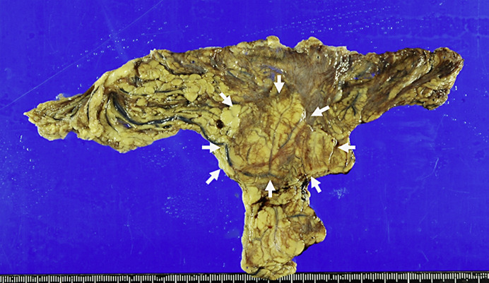
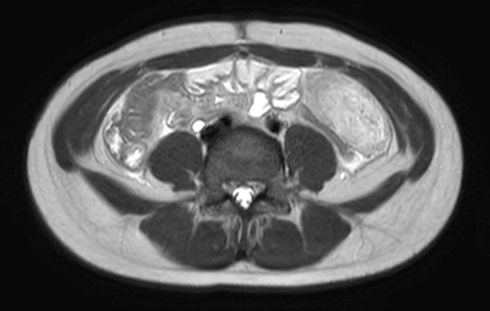
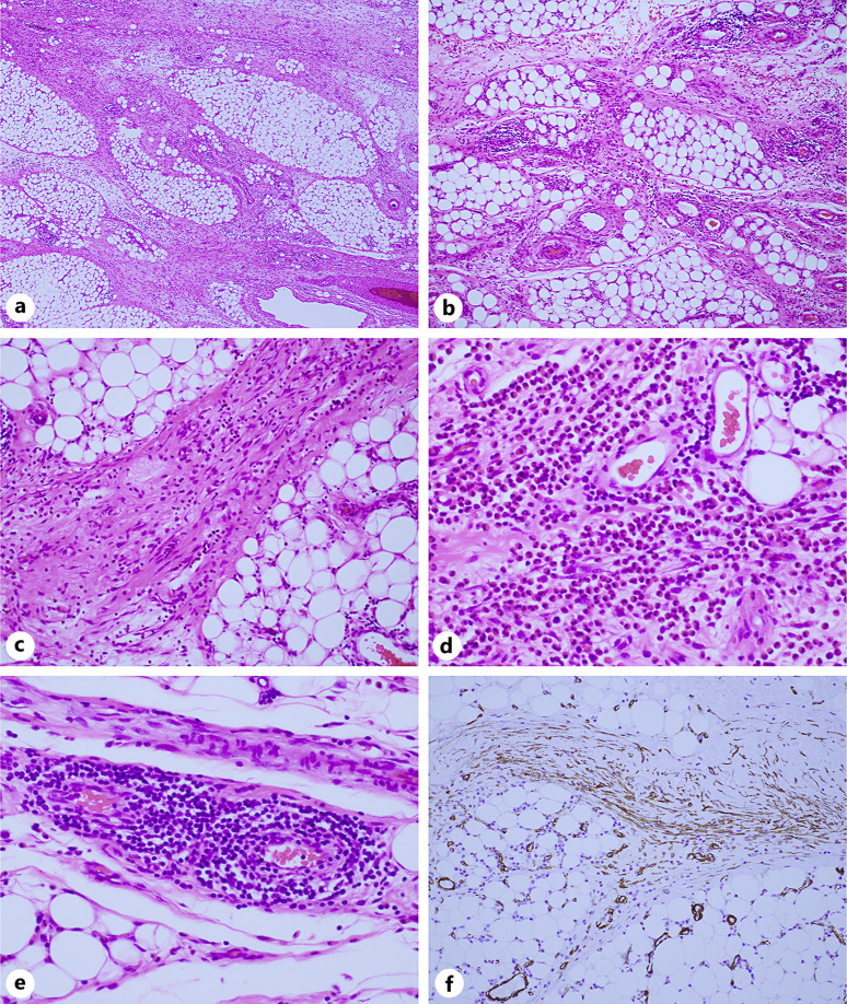
Case presentation: The patient, an 11-year-old girl, complained of dull periumbilical pain and nausea, and radiological examination showed a mass lesion in the abdomino-pelvic cavity. On laparoscopy, a plaque-like, flat mass was seen in the greater omentum, and laparoscopic omental resection was performed. On histopathological examination, the interlobular fibrous septa of omental adipose tissue were widened by inflammatory edema, prominent infiltration of eosinophils, and loose proliferation of myofibroblasts. Dense lymphocytic infiltration was also noted around small veins. Inflammatory changes were mild in the fat lobules, and fat necrosis and infiltration of lipid-laden macrophages were absent. Findings of obliterative phlebitis or arteritis were not seen.
Conclusion: Isolated involvement of the omentum by a panniculitic process is rare, and the pathogenesis of eosinophilic septal panniculitis found in the present case remains unknown, but involvement of a hypersensitivity reaction against some unknown stimuli is presumed, based on the histopathological resemblance of the omental lesions to erythema nodosum or eosinophilic panniculitis of the skin. We should keep in mind the possibility that the omental lesion in this patient is a harbinger of more serious immunological disorders. Careful, long-term follow-up and monitoring of the patient are needed.


 求助内容:
求助内容: 应助结果提醒方式:


