Amanda Freitas da Rosa, Dayana Mara Silva Chaves, Luiz Carlos de Lima Dias-Junior, Gabriela Pasqualin Ghidini, Julia Menezes Savaris, Rayssa Sabino da Silva, Roberta Pinto Pereira, Eduardo Antunes Bortoluzzi, Cleonice da Silveira Teixeira, Lucas da Fonseca Roberti Garcia
{"title":"Effectiveness of supplementary protocols for filling material removal after sealer ultrasonic activation - a laboratory investigation.","authors":"Amanda Freitas da Rosa, Dayana Mara Silva Chaves, Luiz Carlos de Lima Dias-Junior, Gabriela Pasqualin Ghidini, Julia Menezes Savaris, Rayssa Sabino da Silva, Roberta Pinto Pereira, Eduardo Antunes Bortoluzzi, Cleonice da Silveira Teixeira, Lucas da Fonseca Roberti Garcia","doi":"10.1590/1807-3107bor-2025.vol39.034","DOIUrl":null,"url":null,"abstract":"<p><p>Ultrasonic activation of the endodontic sealer makes it difficult to remove the material during endodontic reintervention. Therefore, supplementary removal protocols should be tested to optimize the removal of the remaining filling material. This study assessed the effectiveness of supplementary protocols for filling material removal after sealer ultrasonic activation (UA). Sixty teeth were prepared and distributed into two groups: UA and No UA of the sealer before obturation. Teeth were re-instrumented and two supplementary removal protocols were tested, resulting in six groups (n = 10): NoUA; NoUA+XP (XP-endo Finisher); NoUA+CS (Clearsonic-R1); UA; UA+XP; and UA+CS. Root canals were analyzed under stereomicroscopy and scanning electron microscopy for quantification of the remaining filling material. Considering the total root canal area, the NoUA+CS group had the lowest remaining filling material compared to NoUA+XP, UA+XP and UA+CS groups (p < 0.05). When the root thirds were compared, there was no statistical difference among groups (p > 0.05). The XP-endo Finisher instrument demonstrated the lowest effectiveness when used as a supplementary step. In contrast, the Clearsonic-R1 insert exhibited the highest performance.</p>","PeriodicalId":9240,"journal":{"name":"Brazilian oral research","volume":"39 ","pages":"e034"},"PeriodicalIF":1.3000,"publicationDate":"2025-04-04","publicationTypes":"Journal Article","fieldsOfStudy":null,"isOpenAccess":false,"openAccessPdf":"https://www.ncbi.nlm.nih.gov/pmc/articles/PMC11970515/pdf/","citationCount":"0","resultStr":null,"platform":"Semanticscholar","paperid":null,"PeriodicalName":"Brazilian oral research","FirstCategoryId":"3","ListUrlMain":"https://doi.org/10.1590/1807-3107bor-2025.vol39.034","RegionNum":4,"RegionCategory":"医学","ArticlePicture":[],"TitleCN":null,"AbstractTextCN":null,"PMCID":null,"EPubDate":"2025/1/1 0:00:00","PubModel":"eCollection","JCR":"Q3","JCRName":"DENTISTRY, ORAL SURGERY & MEDICINE","Score":null,"Total":0}
引用次数: 0
Abstract
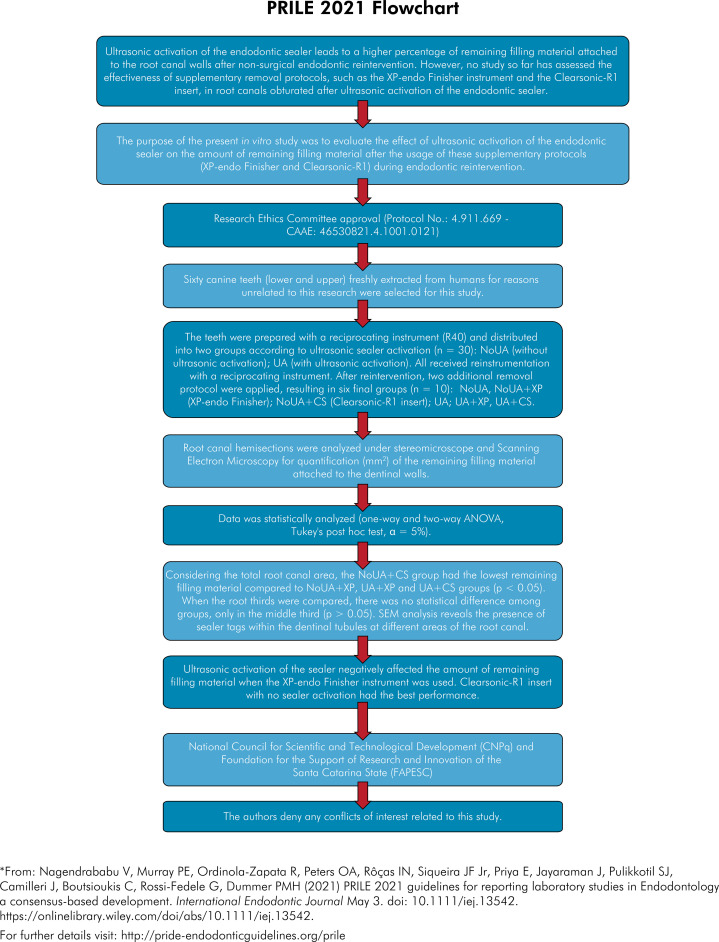
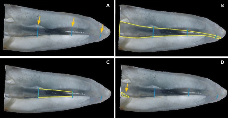
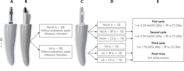
Ultrasonic activation of the endodontic sealer makes it difficult to remove the material during endodontic reintervention. Therefore, supplementary removal protocols should be tested to optimize the removal of the remaining filling material. This study assessed the effectiveness of supplementary protocols for filling material removal after sealer ultrasonic activation (UA). Sixty teeth were prepared and distributed into two groups: UA and No UA of the sealer before obturation. Teeth were re-instrumented and two supplementary removal protocols were tested, resulting in six groups (n = 10): NoUA; NoUA+XP (XP-endo Finisher); NoUA+CS (Clearsonic-R1); UA; UA+XP; and UA+CS. Root canals were analyzed under stereomicroscopy and scanning electron microscopy for quantification of the remaining filling material. Considering the total root canal area, the NoUA+CS group had the lowest remaining filling material compared to NoUA+XP, UA+XP and UA+CS groups (p < 0.05). When the root thirds were compared, there was no statistical difference among groups (p > 0.05). The XP-endo Finisher instrument demonstrated the lowest effectiveness when used as a supplementary step. In contrast, the Clearsonic-R1 insert exhibited the highest performance.


 求助内容:
求助内容: 应助结果提醒方式:


