Density-dependent colour scanning electron microscopy (DDC-SEM). Applications in the study of calcified tissues and visual impact

IF 2.2 3区 工程技术 Q1 MICROSCOPY
Elena Tsolaki , Luke Hunter , Adrian H. Chester , Sergio Bertazzo
{"title":"Density-dependent colour scanning electron microscopy (DDC-SEM). Applications in the study of calcified tissues and visual impact","authors":"Elena Tsolaki ,&nbsp;Luke Hunter ,&nbsp;Adrian H. Chester ,&nbsp;Sergio Bertazzo","doi":"10.1016/j.micron.2025.103821","DOIUrl":null,"url":null,"abstract":"<div><div>Scanning Electron Microscopy (SEM) is widely used as a technique for materials characterization. It has also been successfully applied to the imaging of biological samples, providing invaluable insights into the topography, morphology and composition of biological structures. A particular method combining different SEM detectors, named Density-Dependent Coloured SEM (DDC-SEM), has proven to be most useful for the identification and visualization of minerals in soft tissues. The method consists of a manipulation of original greyscale SEM images to produce coloured images that provide both topography and density information for samples with components of different densities. Here we provide a discussion on how to use DDC-SEM to aid the visualization and intuitive understanding of pathological calcification. This method has become popular not only for its scientific improvement of conventional SEM greyscale images, but also for its aesthetical merits.</div></div>","PeriodicalId":18501,"journal":{"name":"Micron","volume":"195 ","pages":"Article 103821"},"PeriodicalIF":2.2000,"publicationDate":"2025-04-01","publicationTypes":"Journal Article","fieldsOfStudy":null,"isOpenAccess":false,"openAccessPdf":"","citationCount":"0","resultStr":null,"platform":"Semanticscholar","paperid":null,"PeriodicalName":"Micron","FirstCategoryId":"5","ListUrlMain":"https://www.sciencedirect.com/science/article/pii/S0968432825000393","RegionNum":3,"RegionCategory":"工程技术","ArticlePicture":[],"TitleCN":null,"AbstractTextCN":null,"PMCID":null,"EPubDate":"","PubModel":"","JCR":"Q1","JCRName":"MICROSCOPY","Score":null,"Total":0}
引用次数: 0

Abstract

Scanning Electron Microscopy (SEM) is widely used as a technique for materials characterization. It has also been successfully applied to the imaging of biological samples, providing invaluable insights into the topography, morphology and composition of biological structures. A particular method combining different SEM detectors, named Density-Dependent Coloured SEM (DDC-SEM), has proven to be most useful for the identification and visualization of minerals in soft tissues. The method consists of a manipulation of original greyscale SEM images to produce coloured images that provide both topography and density information for samples with components of different densities. Here we provide a discussion on how to use DDC-SEM to aid the visualization and intuitive understanding of pathological calcification. This method has become popular not only for its scientific improvement of conventional SEM greyscale images, but also for its aesthetical merits.
密度依赖性彩色扫描电子显微镜(DDC-SEM)。在钙化组织和视觉冲击研究中的应用
扫描电子显微镜(SEM)是一种广泛应用于材料表征的技术。它还成功地应用于生物样品的成像,为生物结构的地形、形态和组成提供了宝贵的见解。一种特殊的方法结合不同的扫描电镜探测器,称为密度依赖性彩色扫描电镜(DDC-SEM),已被证明是最有用的鉴定和可视化的矿物在软组织。该方法包括对原始灰度扫描电镜图像的操作,以产生彩色图像,为具有不同密度成分的样品提供地形和密度信息。在这里,我们讨论了如何使用DDC-SEM来帮助可视化和直观地理解病理性钙化。该方法不仅对传统的扫描电镜灰度图像进行了科学的改进,而且具有美观的优点。
本文章由计算机程序翻译,如有差异,请以英文原文为准。
求助全文
约1分钟内获得全文 求助全文
来源期刊
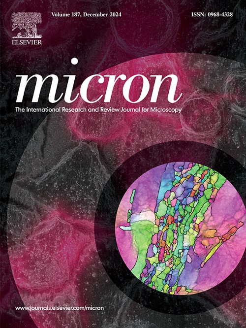
Micron
Micron 工程技术-显微镜技术
CiteScore
4.30
自引率
4.20%
发文量
100
审稿时长
31 days
期刊介绍: Micron is an interdisciplinary forum for all work that involves new applications of microscopy or where advanced microscopy plays a central role. The journal will publish on the design, methods, application, practice or theory of microscopy and microanalysis, including reports on optical, electron-beam, X-ray microtomography, and scanning-probe systems. It also aims at the regular publication of review papers, short communications, as well as thematic issues on contemporary developments in microscopy and microanalysis. The journal embraces original research in which microscopy has contributed significantly to knowledge in biology, life science, nanoscience and nanotechnology, materials science and engineering.
×
引用
GB/T 7714-2015
复制
MLA
复制
APA
复制
导出至
BibTeX EndNote RefMan NoteFirst NoteExpress
×
提示
您的信息不完整,为了账户安全,请先补充。
现在去补充
×
提示
您因"违规操作"
具体请查看互助需知
我知道了
×
提示
确定
请完成安全验证×
copy
已复制链接
快去分享给好友吧!
我知道了
右上角分享
点击右上角分享
0
联系我们:info@booksci.cn Book学术提供免费学术资源搜索服务,方便国内外学者检索中英文文献。致力于提供最便捷和优质的服务体验。 Copyright © 2023 布克学术 All rights reserved.
京ICP备2023020795号-1
ghs 京公网安备 11010802042870号
Book学术文献互助
Book学术文献互助群
群 号:604180095
Book学术官方微信