Rosalba De Sarro, Nunzia Borrelli, Giulia Pelaia, Alessia Mendicino, Sara Moscatelli, Isabella Leo, Giulia La Vecchia, Giuseppe Mazza, Lucy Castaldo, Antonio Strangio, Martina Avesani, Salvatore De Rosa, Daniele Torella, Giovanni Di Salvo, Jolanda Sabatino
{"title":"How to behave with paediatric myocarditis: imaging methods and clinical considerations.","authors":"Rosalba De Sarro, Nunzia Borrelli, Giulia Pelaia, Alessia Mendicino, Sara Moscatelli, Isabella Leo, Giulia La Vecchia, Giuseppe Mazza, Lucy Castaldo, Antonio Strangio, Martina Avesani, Salvatore De Rosa, Daniele Torella, Giovanni Di Salvo, Jolanda Sabatino","doi":"10.1093/ehjimp/qyaf025","DOIUrl":null,"url":null,"abstract":"<p><p>Paediatric myocarditis is a challenging and heterogeneous condition, with varied clinical presentations ranging from mild symptoms to life-threatening complications such as heart failure, arrhythmias, and sudden cardiac death. Effective management hinges on early diagnosis, appropriate treatment, and ongoing monitoring, which can be significantly enhanced through multimodal imaging techniques. This review emphasizes the crucial role of advanced imaging in the diagnosis, prognostication, and management of paediatric myocarditis. While traditional echocardiography remains the first-line imaging tool, it is often insufficient in detecting subtle myocardial changes and it does not allow the identification of myocardial inflammation and fibrosis, particularly in cases with preserved left ventricular function. Recent advancements, including speckle-tracking echocardiography, provide enhanced sensitivity for detecting early signs of myocardial dysfunction, even in the absence of overt abnormalities. Cardiovascular magnetic resonance (CMR) has emerged as a cornerstone in the non-invasive evaluation of myocarditis, offering unparalleled tissue characterization. Indeed, CMR provides critical insights into myocardial oedema, necrosis, and fibrosis, which are essential for confirming the diagnosis, stratifying prognosis, and guiding therapy. Parametric mapping techniques allow for highly accurate detection of myocardial fibrosis (native T1 mapping) and inflammation (T2 mapping), even in the absence of gadolinium contrast, making it particularly valuable in paediatric patients. In conclusion, multimodality imaging, integrating echocardiography and CMR, allows for a comprehensive understanding of disease severity, guiding treatment decisions and optimizing long-term outcomes. This review underscores the importance of a tailored, imaging-driven approach to managing paediatric myocarditis, ensuring the best possible care for this special population.</p>","PeriodicalId":94317,"journal":{"name":"European heart journal. Imaging methods and practice","volume":"3 1","pages":"qyaf025"},"PeriodicalIF":0.0000,"publicationDate":"2025-04-04","publicationTypes":"Journal Article","fieldsOfStudy":null,"isOpenAccess":false,"openAccessPdf":"https://www.ncbi.nlm.nih.gov/pmc/articles/PMC11969066/pdf/","citationCount":"0","resultStr":null,"platform":"Semanticscholar","paperid":null,"PeriodicalName":"European heart journal. Imaging methods and practice","FirstCategoryId":"1085","ListUrlMain":"https://doi.org/10.1093/ehjimp/qyaf025","RegionNum":0,"RegionCategory":null,"ArticlePicture":[],"TitleCN":null,"AbstractTextCN":null,"PMCID":null,"EPubDate":"2025/1/1 0:00:00","PubModel":"eCollection","JCR":"","JCRName":"","Score":null,"Total":0}
引用次数: 0
Abstract
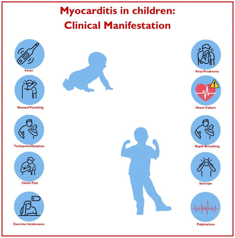
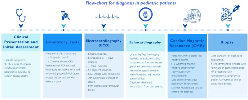
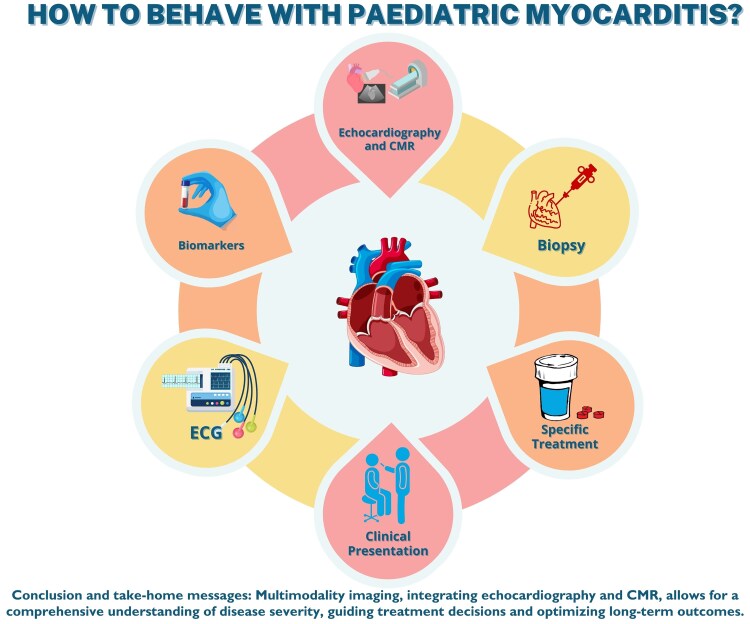
Paediatric myocarditis is a challenging and heterogeneous condition, with varied clinical presentations ranging from mild symptoms to life-threatening complications such as heart failure, arrhythmias, and sudden cardiac death. Effective management hinges on early diagnosis, appropriate treatment, and ongoing monitoring, which can be significantly enhanced through multimodal imaging techniques. This review emphasizes the crucial role of advanced imaging in the diagnosis, prognostication, and management of paediatric myocarditis. While traditional echocardiography remains the first-line imaging tool, it is often insufficient in detecting subtle myocardial changes and it does not allow the identification of myocardial inflammation and fibrosis, particularly in cases with preserved left ventricular function. Recent advancements, including speckle-tracking echocardiography, provide enhanced sensitivity for detecting early signs of myocardial dysfunction, even in the absence of overt abnormalities. Cardiovascular magnetic resonance (CMR) has emerged as a cornerstone in the non-invasive evaluation of myocarditis, offering unparalleled tissue characterization. Indeed, CMR provides critical insights into myocardial oedema, necrosis, and fibrosis, which are essential for confirming the diagnosis, stratifying prognosis, and guiding therapy. Parametric mapping techniques allow for highly accurate detection of myocardial fibrosis (native T1 mapping) and inflammation (T2 mapping), even in the absence of gadolinium contrast, making it particularly valuable in paediatric patients. In conclusion, multimodality imaging, integrating echocardiography and CMR, allows for a comprehensive understanding of disease severity, guiding treatment decisions and optimizing long-term outcomes. This review underscores the importance of a tailored, imaging-driven approach to managing paediatric myocarditis, ensuring the best possible care for this special population.


 求助内容:
求助内容: 应助结果提醒方式:


