Villeseveri Somerkivi, Thorsten Sellerer, Daniel Berthe, York Haemisch, Tuomas Pantsar, Henrik Lohman, Touko Kaasalainen, Franz Pfeiffer
{"title":"Mandible bone mineral density estimation using spectral panoramic X-ray imaging.","authors":"Villeseveri Somerkivi, Thorsten Sellerer, Daniel Berthe, York Haemisch, Tuomas Pantsar, Henrik Lohman, Touko Kaasalainen, Franz Pfeiffer","doi":"10.5624/isd.20240231","DOIUrl":null,"url":null,"abstract":"<p><strong>Purpose: </strong>This study demonstrated the feasibility of obtaining mandible bone mineral density (BMD) scores using spectral panoramic imaging.</p><p><strong>Materials and methods: </strong>Areal BMD scores were measured from the body and angle of the mandible in 3 anthropomorphic head phantoms using a spectral panoramic system (Planmeca Promax Mid, Planmeca Oy, Helsinki, Finland) equipped with a DC-Vela detector (Varex Imaging Corporation, Salt Lake City, USA). These results were compared to synthetic panoramic images generated from dual-energy CT acquisitions. Reproducibility was evaluated by repeatedly scanning 1 phantom with minor patient positioning errors, and the linearity of the BMD scores was assessed using calcium inserts in a Gammex 472 phantom (Sun Nuclear, Melbourne, USA).</p><p><strong>Results: </strong>The experimental and synthetic panoramic images appeared visually similar. The mean synthetic score was 0.640 g/cm<sup>2</sup>, and the anthropomorphic phantoms produced a root mean squared error of 0.0292 g/cm<sup>2</sup> with a correlation coefficient of 0.969. Typical patient positioning errors did not substantially increase the error, which measured 0.0296 g/cm<sup>2</sup> and 0.0474 g/cm<sup>2</sup> for the left and right sides, respectively. Linearity tests using the Gammex phantom yielded a correlation coefficient of 0.998 for BMD scores ranging from 0.03 to 2.7 g/cm<sup>2</sup>.</p><p><strong>Conclusion: </strong>The BMD data obtained from spectral panoramic imaging are consistent with both dual-energy CT and Gammex phantom measurements. Consequently, spectral panoramic imaging shows potential as a method for osteoporosis screening, leveraging the widespread use of panoramic imaging.</p>","PeriodicalId":51714,"journal":{"name":"Imaging Science in Dentistry","volume":"55 1","pages":"56-64"},"PeriodicalIF":2.1000,"publicationDate":"2025-03-01","publicationTypes":"Journal Article","fieldsOfStudy":null,"isOpenAccess":false,"openAccessPdf":"https://www.ncbi.nlm.nih.gov/pmc/articles/PMC11966022/pdf/","citationCount":"0","resultStr":null,"platform":"Semanticscholar","paperid":null,"PeriodicalName":"Imaging Science in Dentistry","FirstCategoryId":"1085","ListUrlMain":"https://doi.org/10.5624/isd.20240231","RegionNum":0,"RegionCategory":null,"ArticlePicture":[],"TitleCN":null,"AbstractTextCN":null,"PMCID":null,"EPubDate":"2025/2/18 0:00:00","PubModel":"Epub","JCR":"Q3","JCRName":"DENTISTRY, ORAL SURGERY & MEDICINE","Score":null,"Total":0}
引用次数: 0
Abstract
Purpose: This study demonstrated the feasibility of obtaining mandible bone mineral density (BMD) scores using spectral panoramic imaging.
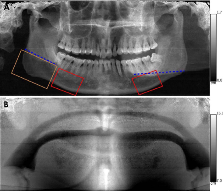
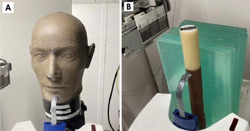
Materials and methods: Areal BMD scores were measured from the body and angle of the mandible in 3 anthropomorphic head phantoms using a spectral panoramic system (Planmeca Promax Mid, Planmeca Oy, Helsinki, Finland) equipped with a DC-Vela detector (Varex Imaging Corporation, Salt Lake City, USA). These results were compared to synthetic panoramic images generated from dual-energy CT acquisitions. Reproducibility was evaluated by repeatedly scanning 1 phantom with minor patient positioning errors, and the linearity of the BMD scores was assessed using calcium inserts in a Gammex 472 phantom (Sun Nuclear, Melbourne, USA).
Results: The experimental and synthetic panoramic images appeared visually similar. The mean synthetic score was 0.640 g/cm2, and the anthropomorphic phantoms produced a root mean squared error of 0.0292 g/cm2 with a correlation coefficient of 0.969. Typical patient positioning errors did not substantially increase the error, which measured 0.0296 g/cm2 and 0.0474 g/cm2 for the left and right sides, respectively. Linearity tests using the Gammex phantom yielded a correlation coefficient of 0.998 for BMD scores ranging from 0.03 to 2.7 g/cm2.
Conclusion: The BMD data obtained from spectral panoramic imaging are consistent with both dual-energy CT and Gammex phantom measurements. Consequently, spectral panoramic imaging shows potential as a method for osteoporosis screening, leveraging the widespread use of panoramic imaging.


 求助内容:
求助内容: 应助结果提醒方式:


