Impact of an intraoral X-ray cone with positioning laser lights on reducing radiographic errors with the bisecting angle technique: A technical report.
{"title":"Impact of an intraoral X-ray cone with positioning laser lights on reducing radiographic errors with the bisecting angle technique: A technical report.","authors":"Suresh Kandagal Veerabhadrappa, Jayanth Kumar Vadivel, Seema Yadav Roodmal, Ummul Khairah Bt Ismail, Vipin Kailasmal Jain","doi":"10.5624/isd.20240196","DOIUrl":null,"url":null,"abstract":"<p><strong>Purpose: </strong>The bisecting angle technique (BAT) encounters difficulties in positioning the X-ray cone and aligning the central beam with the tooth. To address this, a rectangular laser featuring a pointed light was integrated into the intraoral X-ray cone. This study evaluated its effectiveness in improving radiograph quality and minimizing errors.</p><p><strong>Materials and methods: </strong>Fifty fifth-year Bachelor of Dental Surgery students were divided into 3 groups. Group 1 (n=16) used the paralleling technique, group 2 (n=17) employed the conventional BAT, and group 3 (n=17) utilized the laser-assisted BAT on mannequins. Two independent oral radiologists assessed the quality of the radiographs, categorizing the images as either diagnostically acceptable or not acceptable. Inter-group comparisons of quality and error rates were conducted using the chi-square test (significance level: <i>P</i><0.05).</p><p><strong>Results: </strong>The paralleling technique group produced 77.5% diagnostically acceptable radiographs and 22.5% that were not diagnostically acceptable. These percentages were 65.3% and 34.7%, respectively, in the conventional BAT group and 75.3% and 24.7%, respectively, in the laser-assisted BAT group, showing results similar to the paralleling technique group. The quality of radiographs differed significantly among the groups (<i>P</i><0.05). The percentage of error-free radiographs was 38.1% in the paralleling technique group, 20.6% in the conventional BAT group, and 40.0% in the laser-assisted BAT group, with these differences being statistically significant (<i>P</i><0.05).</p><p><strong>Conclusion: </strong>The device produced higher acceptability and fewer radiographic errors than the conventional BAT technique, suggesting accurate adjustment of the X-ray cone and central beam to the desired teeth.</p>","PeriodicalId":51714,"journal":{"name":"Imaging Science in Dentistry","volume":"55 1","pages":"65-71"},"PeriodicalIF":2.1000,"publicationDate":"2025-03-01","publicationTypes":"Journal Article","fieldsOfStudy":null,"isOpenAccess":false,"openAccessPdf":"https://www.ncbi.nlm.nih.gov/pmc/articles/PMC11966014/pdf/","citationCount":"0","resultStr":null,"platform":"Semanticscholar","paperid":null,"PeriodicalName":"Imaging Science in Dentistry","FirstCategoryId":"1085","ListUrlMain":"https://doi.org/10.5624/isd.20240196","RegionNum":0,"RegionCategory":null,"ArticlePicture":[],"TitleCN":null,"AbstractTextCN":null,"PMCID":null,"EPubDate":"2025/1/15 0:00:00","PubModel":"Epub","JCR":"Q3","JCRName":"DENTISTRY, ORAL SURGERY & MEDICINE","Score":null,"Total":0}
引用次数: 0
Abstract
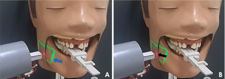
Purpose: The bisecting angle technique (BAT) encounters difficulties in positioning the X-ray cone and aligning the central beam with the tooth. To address this, a rectangular laser featuring a pointed light was integrated into the intraoral X-ray cone. This study evaluated its effectiveness in improving radiograph quality and minimizing errors.
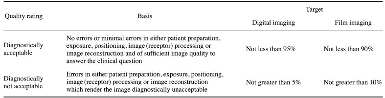
Materials and methods: Fifty fifth-year Bachelor of Dental Surgery students were divided into 3 groups. Group 1 (n=16) used the paralleling technique, group 2 (n=17) employed the conventional BAT, and group 3 (n=17) utilized the laser-assisted BAT on mannequins. Two independent oral radiologists assessed the quality of the radiographs, categorizing the images as either diagnostically acceptable or not acceptable. Inter-group comparisons of quality and error rates were conducted using the chi-square test (significance level: P<0.05).
Results: The paralleling technique group produced 77.5% diagnostically acceptable radiographs and 22.5% that were not diagnostically acceptable. These percentages were 65.3% and 34.7%, respectively, in the conventional BAT group and 75.3% and 24.7%, respectively, in the laser-assisted BAT group, showing results similar to the paralleling technique group. The quality of radiographs differed significantly among the groups (P<0.05). The percentage of error-free radiographs was 38.1% in the paralleling technique group, 20.6% in the conventional BAT group, and 40.0% in the laser-assisted BAT group, with these differences being statistically significant (P<0.05).
Conclusion: The device produced higher acceptability and fewer radiographic errors than the conventional BAT technique, suggesting accurate adjustment of the X-ray cone and central beam to the desired teeth.

 求助内容:
求助内容: 应助结果提醒方式:


