{"title":"Sirtuin 3-mediated delactylation of malic enzyme 2 disrupts redox balance and inhibits colorectal cancer growth.","authors":"Chaoqun Li, Cun Ge, Qingwen Wang, Peng Teng, Heyuan Jia, Surui Yao, Zhaohui Huang","doi":"10.1007/s13402-025-01058-5","DOIUrl":null,"url":null,"abstract":"<p><strong>Purpose: </strong>Post-translational modifications, such as lactylation, are emerging as critical regulators of metabolic enzymes in cancer progression. Mitochondrial malic enzyme 2 (ME2), a key enzyme in the TCA cycle, plays a pivotal role in maintaining redox homeostasis and supporting tumor metabolism. However, the functional significance of ME2 lactylation and its regulatory mechanisms remain unclear. This study investigates the role of ME2 K352 lactylation in modulating enzymatic activity, redox balance, and tumor progression.</p><p><strong>Methods: </strong>Immunoprecipitation and western blotting were used to assess ME2 lactylation and its interaction with Sirtuin 3 (SIRT3). Mass spectrometry identified the lactylation site on ME2. Enzymatic activity was measured using NADH production assays. The functional effects of ME2 K352 lactylation were analyzed by measuring ROS levels, NADP⁺/NADPH ratios, metabolic intermediates, and mitochondrial respiration parameters. Cell proliferation was evaluated via CCK-8 and colony formation assays. Xenograft tumor models and Ki-67 immunohistochemical staining were used to assess tumor growth and proliferation in vivo.</p><p><strong>Results: </strong>Mass spectrometry identified K352 as the primary lactylation site on ME2. Sodium lactate treatment enhanced ME2 lactylation and enzymatic activity, while SIRT3-mediated delactylation at K352 reduced ME2 activity, disrupting redox homeostasis. Cells expressing the K352R mutant exhibited elevated ROS levels, higher NADP⁺/NADPH ratios, and altered levels of metabolic intermediates, including increased malate and lactate with reduced pyruvate. Additionally, re-expression of ME2 K352R in HCT116 cells significantly impaired proliferation and colony formation. In vivo, xenograft models demonstrated that ME2 K352R expression suppressed tumor growth, as evidenced by reduced tumor volume, weight, and Ki-67 staining.</p><p><strong>Conclusions: </strong>This study reveals that ME2 K352 lactylation is a critical regulatory mechanism that modulates enzymatic activity, mitochondrial function, and tumor progression. SIRT3-mediated delactylation of ME2 K352 disrupts redox homeostasis and inhibits tumor growth. These findings highlight the potential of targeting ME2 lactylation as a therapeutic strategy in cancer treatment.</p>","PeriodicalId":49223,"journal":{"name":"Cellular Oncology","volume":" ","pages":"979-990"},"PeriodicalIF":4.8000,"publicationDate":"2025-08-01","publicationTypes":"Journal Article","fieldsOfStudy":null,"isOpenAccess":false,"openAccessPdf":"https://www.ncbi.nlm.nih.gov/pmc/articles/PMC12238175/pdf/","citationCount":"0","resultStr":null,"platform":"Semanticscholar","paperid":null,"PeriodicalName":"Cellular Oncology","FirstCategoryId":"3","ListUrlMain":"https://doi.org/10.1007/s13402-025-01058-5","RegionNum":2,"RegionCategory":"医学","ArticlePicture":[],"TitleCN":null,"AbstractTextCN":null,"PMCID":null,"EPubDate":"2025/4/7 0:00:00","PubModel":"Epub","JCR":"Q2","JCRName":"CELL BIOLOGY","Score":null,"Total":0}
引用次数: 0
Abstract
Purpose: Post-translational modifications, such as lactylation, are emerging as critical regulators of metabolic enzymes in cancer progression. Mitochondrial malic enzyme 2 (ME2), a key enzyme in the TCA cycle, plays a pivotal role in maintaining redox homeostasis and supporting tumor metabolism. However, the functional significance of ME2 lactylation and its regulatory mechanisms remain unclear. This study investigates the role of ME2 K352 lactylation in modulating enzymatic activity, redox balance, and tumor progression.
Methods: Immunoprecipitation and western blotting were used to assess ME2 lactylation and its interaction with Sirtuin 3 (SIRT3). Mass spectrometry identified the lactylation site on ME2. Enzymatic activity was measured using NADH production assays. The functional effects of ME2 K352 lactylation were analyzed by measuring ROS levels, NADP⁺/NADPH ratios, metabolic intermediates, and mitochondrial respiration parameters. Cell proliferation was evaluated via CCK-8 and colony formation assays. Xenograft tumor models and Ki-67 immunohistochemical staining were used to assess tumor growth and proliferation in vivo.
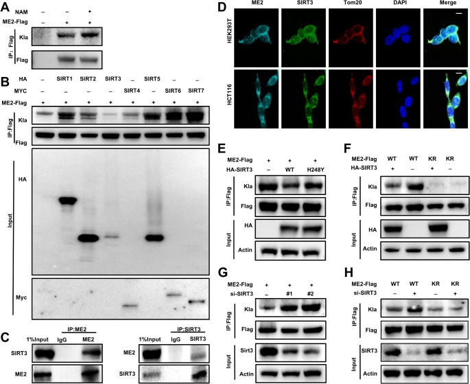
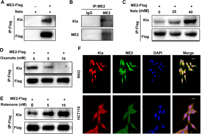
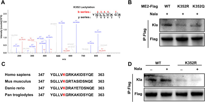
Results: Mass spectrometry identified K352 as the primary lactylation site on ME2. Sodium lactate treatment enhanced ME2 lactylation and enzymatic activity, while SIRT3-mediated delactylation at K352 reduced ME2 activity, disrupting redox homeostasis. Cells expressing the K352R mutant exhibited elevated ROS levels, higher NADP⁺/NADPH ratios, and altered levels of metabolic intermediates, including increased malate and lactate with reduced pyruvate. Additionally, re-expression of ME2 K352R in HCT116 cells significantly impaired proliferation and colony formation. In vivo, xenograft models demonstrated that ME2 K352R expression suppressed tumor growth, as evidenced by reduced tumor volume, weight, and Ki-67 staining.
Conclusions: This study reveals that ME2 K352 lactylation is a critical regulatory mechanism that modulates enzymatic activity, mitochondrial function, and tumor progression. SIRT3-mediated delactylation of ME2 K352 disrupts redox homeostasis and inhibits tumor growth. These findings highlight the potential of targeting ME2 lactylation as a therapeutic strategy in cancer treatment.
期刊介绍:
The Official Journal of the International Society for Cellular Oncology
Focuses on translational research
Addresses the conversion of cell biology to clinical applications
Cellular Oncology publishes scientific contributions from various biomedical and clinical disciplines involved in basic and translational cancer research on the cell and tissue level, technical and bioinformatics developments in this area, and clinical applications. This includes a variety of fields like genome technology, micro-arrays and other high-throughput techniques, genomic instability, SNP, DNA methylation, signaling pathways, DNA organization, (sub)microscopic imaging, proteomics, bioinformatics, functional effects of genomics, drug design and development, molecular diagnostics and targeted cancer therapies, genotype-phenotype interactions.
A major goal is to translate the latest developments in these fields from the research laboratory into routine patient management. To this end Cellular Oncology forms a platform of scientific information exchange between molecular biologists and geneticists, technical developers, pathologists, (medical) oncologists and other clinicians involved in the management of cancer patients.
In vitro studies are preferentially supported by validations in tumor tissue with clinicopathological associations.
 求助内容:
求助内容: 应助结果提醒方式:


