Boyuan Li, Villeseveri Somerkivi, Farhang Bayat, Carolyn Huynh, Cem Altunbas
{"title":"Effect of a prototype 2-dimensional antiscatter grid on image quality obtained with a dental cone-beam computed tomography scanner.","authors":"Boyuan Li, Villeseveri Somerkivi, Farhang Bayat, Carolyn Huynh, Cem Altunbas","doi":"10.5624/isd.20240182","DOIUrl":null,"url":null,"abstract":"<p><strong>Purpose: </strong>X-ray scattering adversely affects cone-beam computed tomography (CBCT) image quality, generating image artifacts, causing inaccurate tissue density representation, and reducing contrast. This study evaluated the performance of a 2-dimensional antiscatter grid (2D grid) prototype in a dental CBCT system.</p><p><strong>Materials and methods: </strong>A focused 2D grid prototype was fabricated from tungsten and integrated with the detector of a dental CBCT system. Residual scatter transmitted through the 2D grid was corrected using a measurement-based scatter correction method. Phantom imaging experiments were performed in anatomical regions relevant to dental and head imaging with and without this grid. Following image reconstruction via filtered back projection, attenuation coefficients were converted to Hounsfield units (HU). Subsequently, scatter suppression performance, HU consistency, image artifacts, and contrast resolution were evaluated.</p><p><strong>Results: </strong>The 2D grid reduced scatter intensity by a factor of 10-20 in CBCT projections. Consequently, the grid substantially increased contrast, reduced image artifacts, and improved HU consistency. The contrast increased by 27% and 48% in bone- and soft tissue-equivalent regions, respectively. HU value deviations among teeth decreased from 510 to 146 HU. These results indicate improved visualization and tissue density representation fidelity in CBCT images acquired with the 2D grid.</p><p><strong>Conclusion: </strong>Use of a 2D grid could substantially improve the accuracy of tissue density representation and the contrast of dental and head anatomy in 3-dimensional images obtained with dental CBCT. Such improvements may translate to better quantitative evaluation of bone quality, enhanced tissue visualization, and more accurate model generation for surgical planning and guidance.</p>","PeriodicalId":51714,"journal":{"name":"Imaging Science in Dentistry","volume":"55 1","pages":"37-47"},"PeriodicalIF":2.1000,"publicationDate":"2025-03-01","publicationTypes":"Journal Article","fieldsOfStudy":null,"isOpenAccess":false,"openAccessPdf":"https://www.ncbi.nlm.nih.gov/pmc/articles/PMC11966021/pdf/","citationCount":"0","resultStr":null,"platform":"Semanticscholar","paperid":null,"PeriodicalName":"Imaging Science in Dentistry","FirstCategoryId":"1085","ListUrlMain":"https://doi.org/10.5624/isd.20240182","RegionNum":0,"RegionCategory":null,"ArticlePicture":[],"TitleCN":null,"AbstractTextCN":null,"PMCID":null,"EPubDate":"2025/2/18 0:00:00","PubModel":"Epub","JCR":"Q3","JCRName":"DENTISTRY, ORAL SURGERY & MEDICINE","Score":null,"Total":0}
引用次数: 0
Abstract
Purpose: X-ray scattering adversely affects cone-beam computed tomography (CBCT) image quality, generating image artifacts, causing inaccurate tissue density representation, and reducing contrast. This study evaluated the performance of a 2-dimensional antiscatter grid (2D grid) prototype in a dental CBCT system.
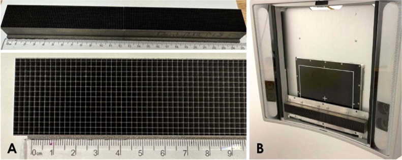
Materials and methods: A focused 2D grid prototype was fabricated from tungsten and integrated with the detector of a dental CBCT system. Residual scatter transmitted through the 2D grid was corrected using a measurement-based scatter correction method. Phantom imaging experiments were performed in anatomical regions relevant to dental and head imaging with and without this grid. Following image reconstruction via filtered back projection, attenuation coefficients were converted to Hounsfield units (HU). Subsequently, scatter suppression performance, HU consistency, image artifacts, and contrast resolution were evaluated.
Results: The 2D grid reduced scatter intensity by a factor of 10-20 in CBCT projections. Consequently, the grid substantially increased contrast, reduced image artifacts, and improved HU consistency. The contrast increased by 27% and 48% in bone- and soft tissue-equivalent regions, respectively. HU value deviations among teeth decreased from 510 to 146 HU. These results indicate improved visualization and tissue density representation fidelity in CBCT images acquired with the 2D grid.
Conclusion: Use of a 2D grid could substantially improve the accuracy of tissue density representation and the contrast of dental and head anatomy in 3-dimensional images obtained with dental CBCT. Such improvements may translate to better quantitative evaluation of bone quality, enhanced tissue visualization, and more accurate model generation for surgical planning and guidance.

 求助内容:
求助内容: 应助结果提醒方式:


