Asmaa T Uthman, Habiba Abouelenen, Shaheer Khan, Omar Bseiso, Natheer Al-Rawi
{"title":"Diagnostic accuracy of artificial intelligence in the detection of maxillary sinus pathology using computed tomography: A concise systematic review.","authors":"Asmaa T Uthman, Habiba Abouelenen, Shaheer Khan, Omar Bseiso, Natheer Al-Rawi","doi":"10.5624/isd.20240139","DOIUrl":null,"url":null,"abstract":"<p><strong>Purpose: </strong>This study was performed to assess the performance and accuracy of artificial intelligence (AI) in the detection and diagnosis of maxillary sinus pathologies using computed tomography (CT)/cone-beam computed tomography (CBCT) imaging.</p><p><strong>Materials and methods: </strong>A comprehensive literature search was conducted across 4 databases: Google Scholar, BioMed Central (BMC), ProQuest, and PubMed. Combinations of keywords such as \"DCNN,\" \"deep learning,\" \"convolutional neural network,\" \"machine learning,\" \"predictive modeling,\" and \"data mining\" were used to identify relevant articles. The study included articles that were published within the last 5 years, written in English, available in full text, and focused on diagnostic accuracy.</p><p><strong>Results: </strong>Of an initial 530 records, 12 studies with a total of 3,349 patients (7,358 images) were included. All articles employed deep learning methods. The most commonly tested pathologies were maxillary rhinosinusitis and maxillary sinusitis, while the most frequently used AI models were convolutional neural network architectures, including ResNet and DenseNet, YOLO, and U-Net. DenseNet and ResNet architectures have demonstrated superior precision in detecting maxillary sinus pathologies due to their capacity to handle deeper networks without overfitting. The performance in detecting maxillary sinus pathology varied, with an accuracy ranging from 85% to 97%, a sensitivity of 87% to 100%, a specificity of 87.2% to 99.7%, and an area under the curve of 0.80 to 0.91.</p><p><strong>Conclusion: </strong>AI with various architectures has been used to detect maxillary sinus abnormalities on CT/CBCT images, achieving near-perfect results. However, further improvements are needed to increase accuracy and consistency.</p>","PeriodicalId":51714,"journal":{"name":"Imaging Science in Dentistry","volume":"55 1","pages":"1-10"},"PeriodicalIF":2.1000,"publicationDate":"2025-03-01","publicationTypes":"Journal Article","fieldsOfStudy":null,"isOpenAccess":false,"openAccessPdf":"https://www.ncbi.nlm.nih.gov/pmc/articles/PMC11966023/pdf/","citationCount":"0","resultStr":null,"platform":"Semanticscholar","paperid":null,"PeriodicalName":"Imaging Science in Dentistry","FirstCategoryId":"1085","ListUrlMain":"https://doi.org/10.5624/isd.20240139","RegionNum":0,"RegionCategory":null,"ArticlePicture":[],"TitleCN":null,"AbstractTextCN":null,"PMCID":null,"EPubDate":"2025/1/15 0:00:00","PubModel":"Epub","JCR":"Q3","JCRName":"DENTISTRY, ORAL SURGERY & MEDICINE","Score":null,"Total":0}
引用次数: 0
Abstract
Purpose: This study was performed to assess the performance and accuracy of artificial intelligence (AI) in the detection and diagnosis of maxillary sinus pathologies using computed tomography (CT)/cone-beam computed tomography (CBCT) imaging.
Materials and methods: A comprehensive literature search was conducted across 4 databases: Google Scholar, BioMed Central (BMC), ProQuest, and PubMed. Combinations of keywords such as "DCNN," "deep learning," "convolutional neural network," "machine learning," "predictive modeling," and "data mining" were used to identify relevant articles. The study included articles that were published within the last 5 years, written in English, available in full text, and focused on diagnostic accuracy.
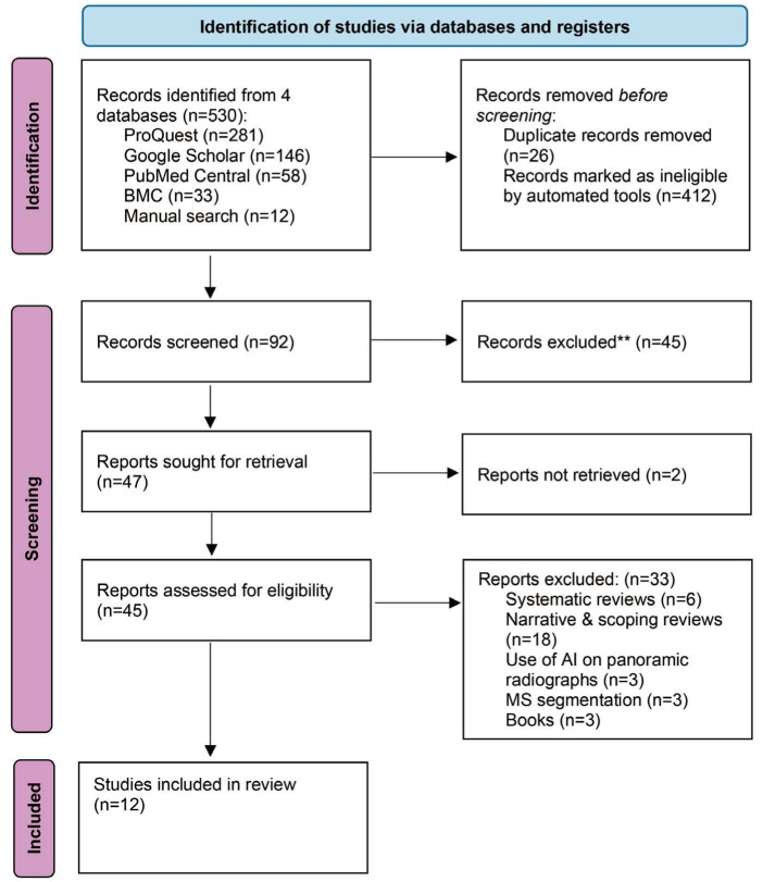
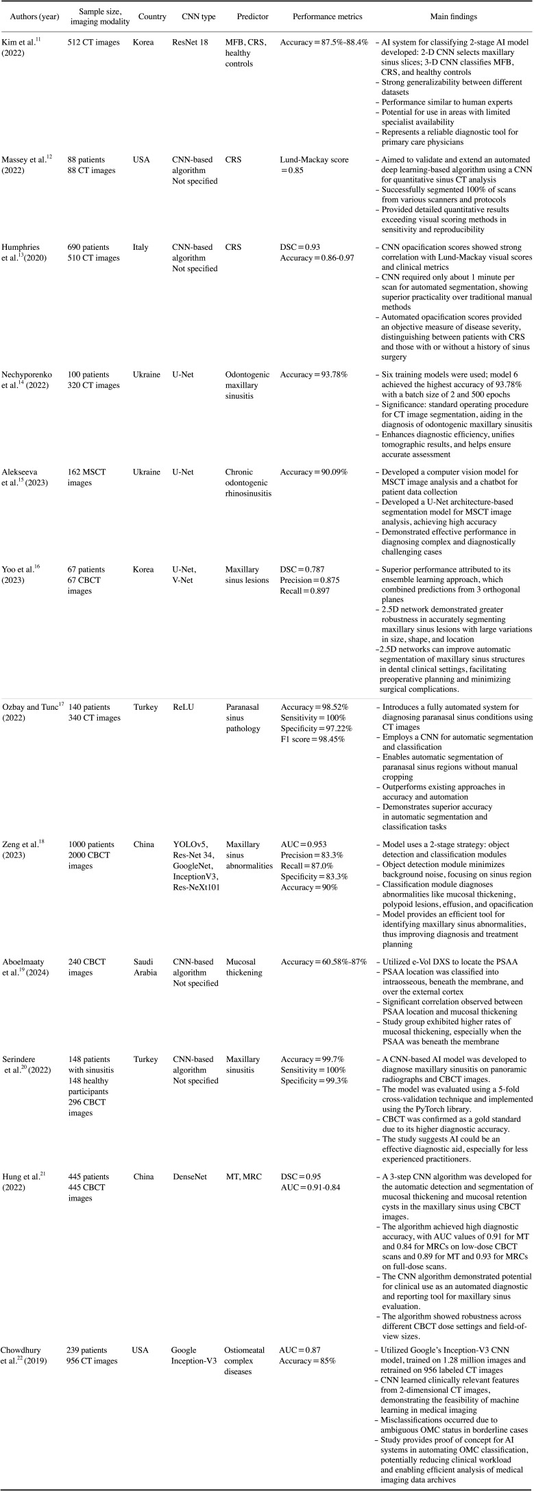
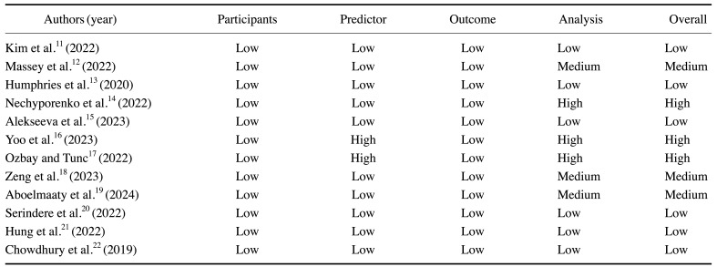
Results: Of an initial 530 records, 12 studies with a total of 3,349 patients (7,358 images) were included. All articles employed deep learning methods. The most commonly tested pathologies were maxillary rhinosinusitis and maxillary sinusitis, while the most frequently used AI models were convolutional neural network architectures, including ResNet and DenseNet, YOLO, and U-Net. DenseNet and ResNet architectures have demonstrated superior precision in detecting maxillary sinus pathologies due to their capacity to handle deeper networks without overfitting. The performance in detecting maxillary sinus pathology varied, with an accuracy ranging from 85% to 97%, a sensitivity of 87% to 100%, a specificity of 87.2% to 99.7%, and an area under the curve of 0.80 to 0.91.
Conclusion: AI with various architectures has been used to detect maxillary sinus abnormalities on CT/CBCT images, achieving near-perfect results. However, further improvements are needed to increase accuracy and consistency.

 求助内容:
求助内容: 应助结果提醒方式:


