Rossano Girometti, Valeria Peruzzi, Paola Clauser, Nina Pötsch, Maria De Martino, Miriam Isola, Gianluca Giannarini, Alessandro Crestani, Chiara Zuiani, Lorenzo Cereser, Pascal At Baltzer
{"title":"Diffusion levels for quantitative assessment of the apparent diffusion coefficient value in prostate MRI: a proof-of-concept bicentric study.","authors":"Rossano Girometti, Valeria Peruzzi, Paola Clauser, Nina Pötsch, Maria De Martino, Miriam Isola, Gianluca Giannarini, Alessandro Crestani, Chiara Zuiani, Lorenzo Cereser, Pascal At Baltzer","doi":"10.1007/s00330-025-11547-8","DOIUrl":null,"url":null,"abstract":"<p><strong>Objectives: </strong>To investigate the performance of Diffusion levels (DLs) in diagnosing clinically significant prostate cancer (csPCa) when combined with the PI-RADS version 2.1.</p><p><strong>Materials and methods: </strong>This retrospective, bicentric study included 261 men who underwent 3.0-T prostate MRI between March 2020 and April 2023, receiving systematic and target prostate biopsy on PI-RADS ≥ 3 lesions. Two readers measured the Apparent diffusion coefficient (ADC) of PI-RADS 1-5 findings in the peripheral zone. By plotting the cumulative frequency of csPCa versus ADCs and using ROC analysis, we derived four DLs expressing levels of restricted diffusion, i.e., very low DL (VL-DL), low DL (L-DL), intermediate DL (I-DL), and high DL (H-DL). We compared the per-lesion diagnostic performance in assessing csPCa (grading group ≥ 2 cancer) assuming to biopsy PI-RADS ≥ 3 lesions (strategy 1), PI-RADS ≥ 3 lesions adjusted with ADC values (strategy 2-4), and PI-RADS ≥ 3 lesions adjusted with DLs (strategy 5-7). Net benefit was assessed with decision curve analysis.</p><p><strong>Results: </strong>csPCa was found in 79/261 men (30.3%) and 152/528 lesions (28.8%). There was a negative correlation (p < 0.0001) between ADC versus malignancy rate (tau -0.970) and DLs versus csPCa grading group (tau -0.614). csPCa prevalence was highest in VL-DL (72.2%) and L-DL (54.4%). Most DLs-based strategies increased specificity, positive predictive value (PPV), and net benefit compared to ADC-based strategies or PI-RADS alone. The best strategy showed 94.7% sensitivity, 82.9% specificity, 69.2% PPV, and 97.5% negative predictive value.</p><p><strong>Conclusion: </strong>While larger-scale validation is needed, DLs have the potential to improve PI-RADS-based biopsy decisions for detecting csPCa in the peripheral zone.</p><p><strong>Key points: </strong>Question It is still unclear how to incorporate quantitative information from diffusion-weighted imaging (DWI) into prostate MRI. Findings Combining DWI-derived diffusion levels (DLs) with the PI-RADS version 2.1 categorization reduced false positives while preserving high sensitivity for clinically significant prostate cancer. Clinical relevance DLs permit to easily account for ADC values of prostate lesions and, in turn, refine biopsy decisions.</p>","PeriodicalId":12076,"journal":{"name":"European Radiology","volume":" ","pages":"6171-6182"},"PeriodicalIF":4.7000,"publicationDate":"2025-10-01","publicationTypes":"Journal Article","fieldsOfStudy":null,"isOpenAccess":false,"openAccessPdf":"https://www.ncbi.nlm.nih.gov/pmc/articles/PMC12417251/pdf/","citationCount":"0","resultStr":null,"platform":"Semanticscholar","paperid":null,"PeriodicalName":"European Radiology","FirstCategoryId":"3","ListUrlMain":"https://doi.org/10.1007/s00330-025-11547-8","RegionNum":2,"RegionCategory":"医学","ArticlePicture":[],"TitleCN":null,"AbstractTextCN":null,"PMCID":null,"EPubDate":"2025/4/7 0:00:00","PubModel":"Epub","JCR":"Q1","JCRName":"RADIOLOGY, NUCLEAR MEDICINE & MEDICAL IMAGING","Score":null,"Total":0}
引用次数: 0
Abstract
Objectives: To investigate the performance of Diffusion levels (DLs) in diagnosing clinically significant prostate cancer (csPCa) when combined with the PI-RADS version 2.1.
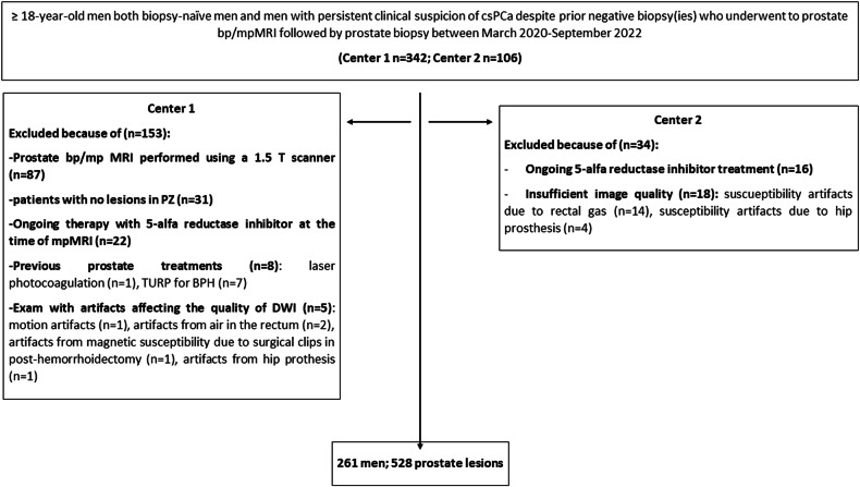
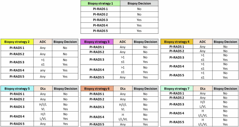
Materials and methods: This retrospective, bicentric study included 261 men who underwent 3.0-T prostate MRI between March 2020 and April 2023, receiving systematic and target prostate biopsy on PI-RADS ≥ 3 lesions. Two readers measured the Apparent diffusion coefficient (ADC) of PI-RADS 1-5 findings in the peripheral zone. By plotting the cumulative frequency of csPCa versus ADCs and using ROC analysis, we derived four DLs expressing levels of restricted diffusion, i.e., very low DL (VL-DL), low DL (L-DL), intermediate DL (I-DL), and high DL (H-DL). We compared the per-lesion diagnostic performance in assessing csPCa (grading group ≥ 2 cancer) assuming to biopsy PI-RADS ≥ 3 lesions (strategy 1), PI-RADS ≥ 3 lesions adjusted with ADC values (strategy 2-4), and PI-RADS ≥ 3 lesions adjusted with DLs (strategy 5-7). Net benefit was assessed with decision curve analysis.
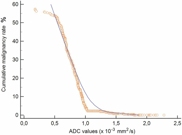
Results: csPCa was found in 79/261 men (30.3%) and 152/528 lesions (28.8%). There was a negative correlation (p < 0.0001) between ADC versus malignancy rate (tau -0.970) and DLs versus csPCa grading group (tau -0.614). csPCa prevalence was highest in VL-DL (72.2%) and L-DL (54.4%). Most DLs-based strategies increased specificity, positive predictive value (PPV), and net benefit compared to ADC-based strategies or PI-RADS alone. The best strategy showed 94.7% sensitivity, 82.9% specificity, 69.2% PPV, and 97.5% negative predictive value.
Conclusion: While larger-scale validation is needed, DLs have the potential to improve PI-RADS-based biopsy decisions for detecting csPCa in the peripheral zone.
Key points: Question It is still unclear how to incorporate quantitative information from diffusion-weighted imaging (DWI) into prostate MRI. Findings Combining DWI-derived diffusion levels (DLs) with the PI-RADS version 2.1 categorization reduced false positives while preserving high sensitivity for clinically significant prostate cancer. Clinical relevance DLs permit to easily account for ADC values of prostate lesions and, in turn, refine biopsy decisions.
期刊介绍:
European Radiology (ER) continuously updates scientific knowledge in radiology by publication of strong original articles and state-of-the-art reviews written by leading radiologists. A well balanced combination of review articles, original papers, short communications from European radiological congresses and information on society matters makes ER an indispensable source for current information in this field.
This is the Journal of the European Society of Radiology, and the official journal of a number of societies.
From 2004-2008 supplements to European Radiology were published under its companion, European Radiology Supplements, ISSN 1613-3749.

 求助内容:
求助内容: 应助结果提醒方式:


