Jae Ang Sim, Sang-Jin Lee, Jung-Min Shin, Byung Hoon Lee
{"title":"Distinct patterns of ligament and meniscal injuries in multiligamentous knee injuries with and without dislocation: a 15-year retrospective study.","authors":"Jae Ang Sim, Sang-Jin Lee, Jung-Min Shin, Byung Hoon Lee","doi":"10.1007/s00068-024-02740-3","DOIUrl":null,"url":null,"abstract":"<p><strong>Purpose: </strong>This study aimed to compare the incidence rates of concomitant injuries, including meniscal and cartilage injuries, between multiligamentous knee injuries (MLKI) with and without dislocation based on our 15-year experience of knee dislocation and MLKI at a level 1 trauma center.</p><p><strong>Methods: </strong>We retrospectively identified 100 patients (115 knees) with MLKIs and/or dislocations at our trauma center between 2007 and 2021. Magnetic resonance imaging was routinely performed to evaluate the injured structures and extent of injury. The anatomic structures of the knee were categorized into anterior and posterior cruciate ligaments (ACL, PCL) and medial and posterolateral structures, and further classified according to the modified Schenck classification. The study participants were divided into two groups: 40 and 75 knees classified as MLKI with and without dislocation, respectively.</p><p><strong>Results: </strong>MLKIs with dislocations showed 13% (5/40 knees) and 18% (7/40 knees) incidence, whereas MLKIs without dislocation showed 15% (11/75 knees) and 13% (10/75 knees) incidence of medial and lateral meniscal tears respectively. The two groups also had a significant discrepancy in the patterns of meniscal tears. For medial meniscal tears, radial tears were more prevalent in MLKIs with dislocation, and longitudinal tears in MLKIs without dislocation (p = 0.197). For lateral meniscal tears, anterior horn or totally detached tears were more prevalent in MLKIs with dislocation, and radial tears in MLKIs without dislocation (p = 0.026). Additionally, complete rupture of all four major ligaments was found in 38% (15/40 knees) of the cases with dislocation, with the majority showing complete ruptures of both the ACL and PCL. Concomitant serious injuries, such as popliteal artery injury and fractures, were observed only in cases involving high-energy trauma and dislocation.</p><p><strong>Conclusions: </strong>MLKIs with dislocation show distinct ligament and meniscal injury patterns compared to those without, highlighting the importance of severity and anatomical classification in diagnosing associated knee injuries.</p><p><strong>Clinical relevance: </strong>The initial distinction in the severity of MLKIs, along with the anatomical classification, have practical implications in identifying associated meniscal tears and injuries to structures surrounding the knee joint.</p><p><strong>Level of evidence: </strong>IV Retrospective comparative study.</p>","PeriodicalId":12064,"journal":{"name":"European Journal of Trauma and Emergency Surgery","volume":"51 1","pages":"163"},"PeriodicalIF":2.2000,"publicationDate":"2025-04-07","publicationTypes":"Journal Article","fieldsOfStudy":null,"isOpenAccess":false,"openAccessPdf":"https://www.ncbi.nlm.nih.gov/pmc/articles/PMC11976805/pdf/","citationCount":"0","resultStr":null,"platform":"Semanticscholar","paperid":null,"PeriodicalName":"European Journal of Trauma and Emergency Surgery","FirstCategoryId":"3","ListUrlMain":"https://doi.org/10.1007/s00068-024-02740-3","RegionNum":3,"RegionCategory":"医学","ArticlePicture":[],"TitleCN":null,"AbstractTextCN":null,"PMCID":null,"EPubDate":"","PubModel":"","JCR":"Q2","JCRName":"EMERGENCY MEDICINE","Score":null,"Total":0}
引用次数: 0
Abstract
Purpose: This study aimed to compare the incidence rates of concomitant injuries, including meniscal and cartilage injuries, between multiligamentous knee injuries (MLKI) with and without dislocation based on our 15-year experience of knee dislocation and MLKI at a level 1 trauma center.
Methods: We retrospectively identified 100 patients (115 knees) with MLKIs and/or dislocations at our trauma center between 2007 and 2021. Magnetic resonance imaging was routinely performed to evaluate the injured structures and extent of injury. The anatomic structures of the knee were categorized into anterior and posterior cruciate ligaments (ACL, PCL) and medial and posterolateral structures, and further classified according to the modified Schenck classification. The study participants were divided into two groups: 40 and 75 knees classified as MLKI with and without dislocation, respectively.
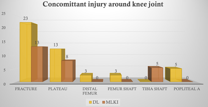
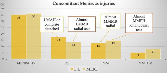
Results: MLKIs with dislocations showed 13% (5/40 knees) and 18% (7/40 knees) incidence, whereas MLKIs without dislocation showed 15% (11/75 knees) and 13% (10/75 knees) incidence of medial and lateral meniscal tears respectively. The two groups also had a significant discrepancy in the patterns of meniscal tears. For medial meniscal tears, radial tears were more prevalent in MLKIs with dislocation, and longitudinal tears in MLKIs without dislocation (p = 0.197). For lateral meniscal tears, anterior horn or totally detached tears were more prevalent in MLKIs with dislocation, and radial tears in MLKIs without dislocation (p = 0.026). Additionally, complete rupture of all four major ligaments was found in 38% (15/40 knees) of the cases with dislocation, with the majority showing complete ruptures of both the ACL and PCL. Concomitant serious injuries, such as popliteal artery injury and fractures, were observed only in cases involving high-energy trauma and dislocation.
Conclusions: MLKIs with dislocation show distinct ligament and meniscal injury patterns compared to those without, highlighting the importance of severity and anatomical classification in diagnosing associated knee injuries.
Clinical relevance: The initial distinction in the severity of MLKIs, along with the anatomical classification, have practical implications in identifying associated meniscal tears and injuries to structures surrounding the knee joint.
Level of evidence: IV Retrospective comparative study.
期刊介绍:
The European Journal of Trauma and Emergency Surgery aims to open an interdisciplinary forum that allows for the scientific exchange between basic and clinical science related to pathophysiology, diagnostics and treatment of traumatized patients. The journal covers all aspects of clinical management, operative treatment and related research of traumatic injuries.
Clinical and experimental papers on issues relevant for the improvement of trauma care are published. Reviews, original articles, short communications and letters allow the appropriate presentation of major and minor topics.
 求助内容:
求助内容: 应助结果提醒方式:


