Yang Wu, Minhui Wang, Chaojun Hu, Shangzhu Zhang, Jiuliang Zhao, Qian Wang, Dong Xu, Xinping Tian, Yan Zhao, Xiaofeng Zeng, Mengtao Li
{"title":"IgG glycosylation profiling of systemic lupus erythematosus using lectin microarray.","authors":"Yang Wu, Minhui Wang, Chaojun Hu, Shangzhu Zhang, Jiuliang Zhao, Qian Wang, Dong Xu, Xinping Tian, Yan Zhao, Xiaofeng Zeng, Mengtao Li","doi":"10.1136/lupus-2024-001413","DOIUrl":null,"url":null,"abstract":"<p><strong>Objectives: </strong>Research on the specific role of immunoglobulin G (IgG) glycosylation in SLE development and progression is limited, especially regarding changes in IgG glycosylation profiles among different SLE subtypes. In this study, we aimed to characterise the glycosylation profile of serum IgG in patients with SLE.</p><p><strong>Methods: </strong>Lectin microarrays with 56 lectins were used to analyse serum IgG glycosylation in 194 patients with SLE, 100 disease controls (40 primary Sjögren's syndrome (pSS), 60 rheumatoid arthritis (RA)) and 100 healthy controls (HCs). Differences between SLE and control groups, as well as SLE subgroups, were validated by lectin blotting. Altered IgG glycosylation patterns were identified and further confirmed. Receiver operating characteristic (ROC) analysis evaluated the diagnostic value of these glycosylation changes in SLE and its subgroups, including neuropsychiatric SLE (NPSLE), lupus nephritis (LN), pulmonary arterial hypertension, immune thrombocytopaenia and SLE without major organ involvement (WMOI).</p><p><strong>Results: </strong>Compared to DC and HC groups, the IgG glycan level of Galβ3GalNAc (binding Jacalin (11.3%) and Maclura pomifera lectin (14.4%)) was significantly increased, whereas most IgG glycan levels were significantly decreased, including core fucose, high mannose, GlcNAc, GalNAc and Galβ4GlcNAc in the SLE group (all p<0.05).The IgG glycan levels were elevated in GalNAc and galactose patterns in the NPSLE group compared to the WMOI group, as well as higher Galβ3GalNAc and galactose patterns in NPSLE and LN compared to HCs.Moreover, ROC curve analysis showed PNA levels might have moderate potential for discriminating SLE from pSS.</p><p><strong>Conclusions: </strong>Patients with SLE show disease-specific alterations in serum IgG glycosylation, and aberrant Galβ3GalNAc, galactose and GalNAc glycosylation may have diagnostic value for SLE and NPSLE. Abnormal IgG glycans may provide new insights into their roles in SLE pathogenesis and progression.</p>","PeriodicalId":18126,"journal":{"name":"Lupus Science & Medicine","volume":"12 1","pages":""},"PeriodicalIF":3.5000,"publicationDate":"2025-04-05","publicationTypes":"Journal Article","fieldsOfStudy":null,"isOpenAccess":false,"openAccessPdf":"https://www.ncbi.nlm.nih.gov/pmc/articles/PMC11973783/pdf/","citationCount":"0","resultStr":null,"platform":"Semanticscholar","paperid":null,"PeriodicalName":"Lupus Science & Medicine","FirstCategoryId":"3","ListUrlMain":"https://doi.org/10.1136/lupus-2024-001413","RegionNum":2,"RegionCategory":"医学","ArticlePicture":[],"TitleCN":null,"AbstractTextCN":null,"PMCID":null,"EPubDate":"","PubModel":"","JCR":"Q1","JCRName":"RHEUMATOLOGY","Score":null,"Total":0}
引用次数: 0
Abstract
Objectives: Research on the specific role of immunoglobulin G (IgG) glycosylation in SLE development and progression is limited, especially regarding changes in IgG glycosylation profiles among different SLE subtypes. In this study, we aimed to characterise the glycosylation profile of serum IgG in patients with SLE.
Methods: Lectin microarrays with 56 lectins were used to analyse serum IgG glycosylation in 194 patients with SLE, 100 disease controls (40 primary Sjögren's syndrome (pSS), 60 rheumatoid arthritis (RA)) and 100 healthy controls (HCs). Differences between SLE and control groups, as well as SLE subgroups, were validated by lectin blotting. Altered IgG glycosylation patterns were identified and further confirmed. Receiver operating characteristic (ROC) analysis evaluated the diagnostic value of these glycosylation changes in SLE and its subgroups, including neuropsychiatric SLE (NPSLE), lupus nephritis (LN), pulmonary arterial hypertension, immune thrombocytopaenia and SLE without major organ involvement (WMOI).
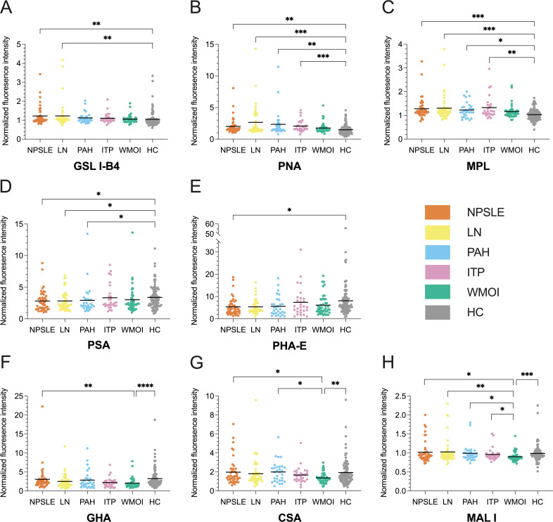
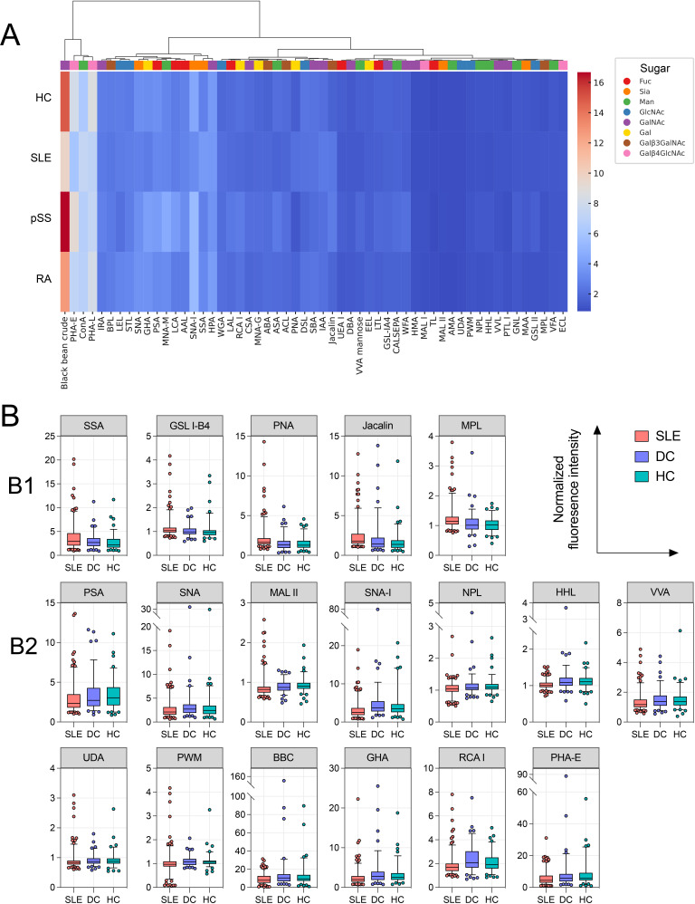
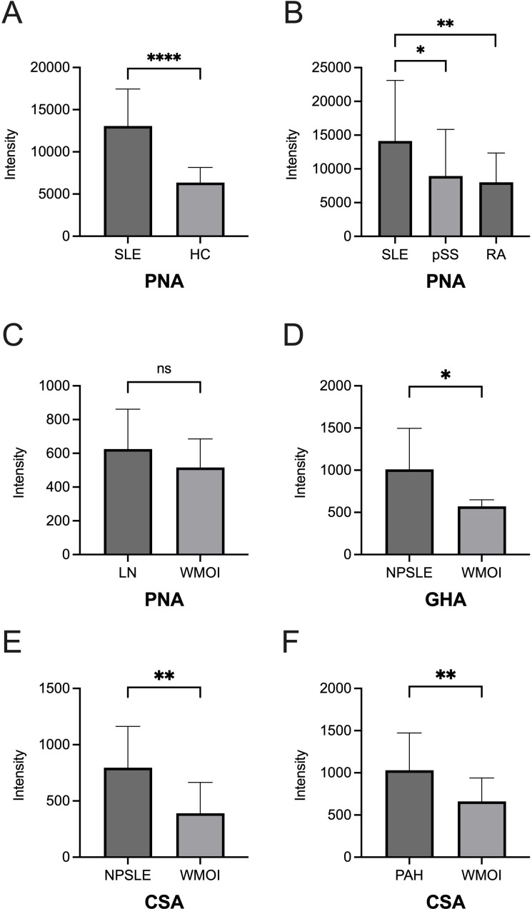
Results: Compared to DC and HC groups, the IgG glycan level of Galβ3GalNAc (binding Jacalin (11.3%) and Maclura pomifera lectin (14.4%)) was significantly increased, whereas most IgG glycan levels were significantly decreased, including core fucose, high mannose, GlcNAc, GalNAc and Galβ4GlcNAc in the SLE group (all p<0.05).The IgG glycan levels were elevated in GalNAc and galactose patterns in the NPSLE group compared to the WMOI group, as well as higher Galβ3GalNAc and galactose patterns in NPSLE and LN compared to HCs.Moreover, ROC curve analysis showed PNA levels might have moderate potential for discriminating SLE from pSS.
Conclusions: Patients with SLE show disease-specific alterations in serum IgG glycosylation, and aberrant Galβ3GalNAc, galactose and GalNAc glycosylation may have diagnostic value for SLE and NPSLE. Abnormal IgG glycans may provide new insights into their roles in SLE pathogenesis and progression.
期刊介绍:
Lupus Science & Medicine is a global, peer reviewed, open access online journal that provides a central point for publication of basic, clinical, translational, and epidemiological studies of all aspects of lupus and related diseases. It is the first lupus-specific open access journal in the world and was developed in response to the need for a barrier-free forum for publication of groundbreaking studies in lupus. The journal publishes research on lupus from fields including, but not limited to: rheumatology, dermatology, nephrology, immunology, pediatrics, cardiology, hepatology, pulmonology, obstetrics and gynecology, and psychiatry.
 求助内容:
求助内容: 应助结果提醒方式:


