Dirk Wiechmann, Robert Leven, Per Rank, Yann Janssens, Jonas Q Schmid
{"title":"Dentoalveolar process remodelling in the anterior mandible after Class III camouflage treatment with lower premolar extractions.","authors":"Dirk Wiechmann, Robert Leven, Per Rank, Yann Janssens, Jonas Q Schmid","doi":"10.1186/s13005-025-00493-x","DOIUrl":null,"url":null,"abstract":"<p><strong>Background: </strong>The aim of this investigation was to evaluate if the hard and soft tissue dentoalveolar process of the mandible follows the tooth movements after lower premolar extractions and anterior retraction in Class III camouflage treatment.</p><p><strong>Methods: </strong>This retrospective study included 25 patients in retention (f/m 12,13) who had previously been treated with lower premolar extractions for Class III camouflage with a completely customized lingual appliance (Wits at T0 -6.7, ± 2.5 mm). The periodontal and dental health of the lower 6 anterior teeth was evaluated (T1) by a thermal sensitivity test, probing and visual inspection after a mean retention period of 3.1 years (± 2.5, min/max 1.0/9.6 years). A novel non-invasive method was used to measure the thickness of the hard and soft tissue dentoalveolar process on the labial and lingual side of the teeth before treatment (T0) and in retention (T1) at 3 different levels using superimposed intraoral scans. A paired t-test with α = 5% was used to evaluate differences between the endpoints.</p><p><strong>Results: </strong>At T1, all 25 patients (mean age 26.8 ± 9.7 years, min/max 16.3/49.5 years) presented uncompromised periodontal and dental situations in the lower anterior segment. The presented digital method for evaluating dimensional changes of the dentoalveolar process had excellent reliability (ICC) with a method error of 0.01 mm. The mean total labio-lingual dimension of the hard and soft tissue dentoalveolar process (min/max 7.89/10.02 mm at T0) was identical at T0 and T1 (mean change of 0.00 ± 0.33 mm, min/max -0.98/0.8 mm). At all levels, the teeth moved only 0.12 mm to the lingual side within the dentoalveolar process and therefore, they moved with the dentoalveolar process and not through it.</p><p><strong>Conclusion: </strong>In non-surgical camouflage treatment with lower premolar extractions in moderate to severe Class III malocclusions, the dentoalveolar process can follow the movement of the mandibular incisors and canines during controlled retraction without any adverse effects.</p>","PeriodicalId":12994,"journal":{"name":"Head & Face Medicine","volume":"21 1","pages":"25"},"PeriodicalIF":2.4000,"publicationDate":"2025-04-04","publicationTypes":"Journal Article","fieldsOfStudy":null,"isOpenAccess":false,"openAccessPdf":"https://www.ncbi.nlm.nih.gov/pmc/articles/PMC11969860/pdf/","citationCount":"0","resultStr":null,"platform":"Semanticscholar","paperid":null,"PeriodicalName":"Head & Face Medicine","FirstCategoryId":"3","ListUrlMain":"https://doi.org/10.1186/s13005-025-00493-x","RegionNum":2,"RegionCategory":"医学","ArticlePicture":[],"TitleCN":null,"AbstractTextCN":null,"PMCID":null,"EPubDate":"","PubModel":"","JCR":"Q2","JCRName":"DENTISTRY, ORAL SURGERY & MEDICINE","Score":null,"Total":0}
引用次数: 0
Abstract
Background: The aim of this investigation was to evaluate if the hard and soft tissue dentoalveolar process of the mandible follows the tooth movements after lower premolar extractions and anterior retraction in Class III camouflage treatment.
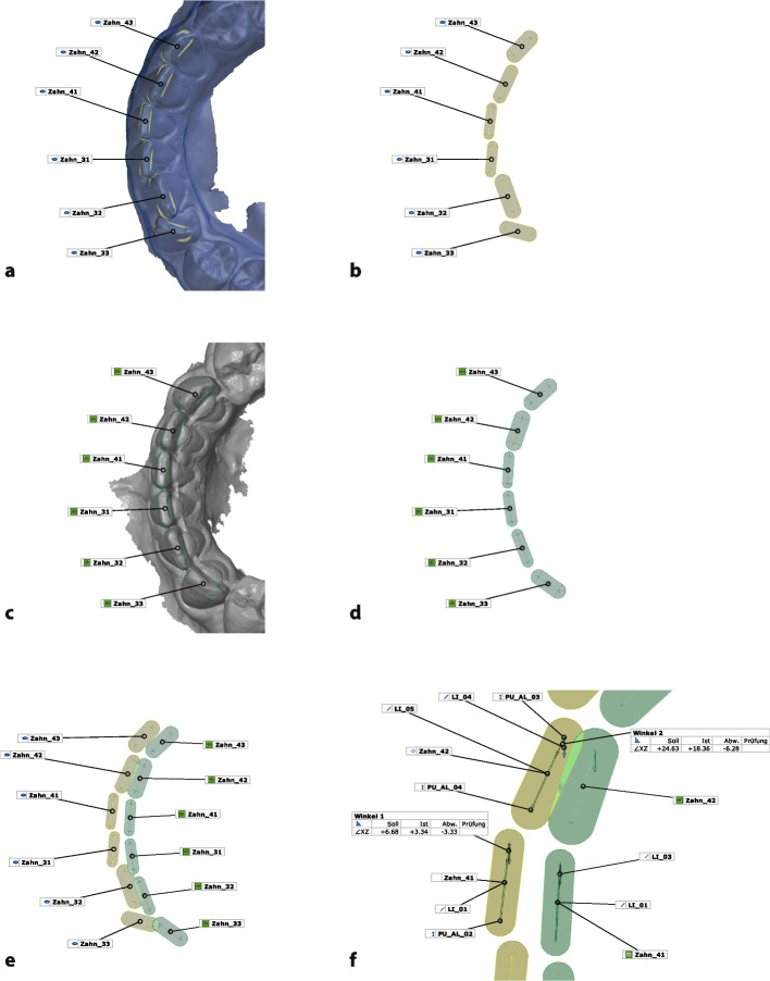
Methods: This retrospective study included 25 patients in retention (f/m 12,13) who had previously been treated with lower premolar extractions for Class III camouflage with a completely customized lingual appliance (Wits at T0 -6.7, ± 2.5 mm). The periodontal and dental health of the lower 6 anterior teeth was evaluated (T1) by a thermal sensitivity test, probing and visual inspection after a mean retention period of 3.1 years (± 2.5, min/max 1.0/9.6 years). A novel non-invasive method was used to measure the thickness of the hard and soft tissue dentoalveolar process on the labial and lingual side of the teeth before treatment (T0) and in retention (T1) at 3 different levels using superimposed intraoral scans. A paired t-test with α = 5% was used to evaluate differences between the endpoints.
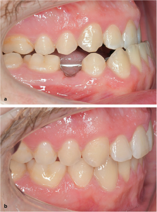
Results: At T1, all 25 patients (mean age 26.8 ± 9.7 years, min/max 16.3/49.5 years) presented uncompromised periodontal and dental situations in the lower anterior segment. The presented digital method for evaluating dimensional changes of the dentoalveolar process had excellent reliability (ICC) with a method error of 0.01 mm. The mean total labio-lingual dimension of the hard and soft tissue dentoalveolar process (min/max 7.89/10.02 mm at T0) was identical at T0 and T1 (mean change of 0.00 ± 0.33 mm, min/max -0.98/0.8 mm). At all levels, the teeth moved only 0.12 mm to the lingual side within the dentoalveolar process and therefore, they moved with the dentoalveolar process and not through it.
Conclusion: In non-surgical camouflage treatment with lower premolar extractions in moderate to severe Class III malocclusions, the dentoalveolar process can follow the movement of the mandibular incisors and canines during controlled retraction without any adverse effects.
期刊介绍:
Head & Face Medicine is a multidisciplinary open access journal that publishes basic and clinical research concerning all aspects of cranial, facial and oral conditions.
The journal covers all aspects of cranial, facial and oral diseases and their management. It has been designed as a multidisciplinary journal for clinicians and researchers involved in the diagnostic and therapeutic aspects of diseases which affect the human head and face. The journal is wide-ranging, covering the development, aetiology, epidemiology and therapy of head and face diseases to the basic science that underlies these diseases. Management of head and face diseases includes all aspects of surgical and non-surgical treatments including psychopharmacological therapies.
 求助内容:
求助内容: 应助结果提醒方式:


