Predictive value of extracellular volume fraction determined using enhanced computed tomography for pathological grading of clear cell renal cell carcinoma: a preliminary study.
{"title":"Predictive value of extracellular volume fraction determined using enhanced computed tomography for pathological grading of clear cell renal cell carcinoma: a preliminary study.","authors":"Jian Liu, Xunlan Zhang, Rui Lv, Xiaoyong Zhang, Rongpin Wang, Xianchun Zeng","doi":"10.1186/s40644-025-00866-0","DOIUrl":null,"url":null,"abstract":"<p><strong>Objective: </strong>To explore the potential of using the extracellular volume fraction (ECV), measured through enhanced computed tomography (CT), as a tool for determining the pathological grade of clear cell renal cell carcinoma (ccRCC).</p><p><strong>Methods: </strong>This retrospective study, approved by the institutional review board, included 65 patients (median age: 58.40 ± 10.84 years) who were diagnosed with ccRCC based on the nucleolar grading of the International Society of Urological Pathology (ISUP). All patients underwent preoperative abdominal enhanced CT between January 2022 and August 2024. CT features from the unenhanced, corticomedullary, nephrographic, and delayed phases were analyzed, and the extracellular volume fraction (ECV) of ccRCC was calculated by measuring CT values from regions of interest in both the unenhanced and nephrographic phases. Statistical significance was evaluated for differences in these parameters across the four ISUP grades. Additionally, diagnostic efficiency was assessed using receiver operating characteristic (ROC) curve analysis.</p><p><strong>Results: </strong>The ECV showed significant differences across the four ISUP grades of ccRCC, its potential as an important predictor of high-grade ccRCC (P = 0.035). The ROC curve analysis indicated that ECV exhibited the highest diagnostic efficacy for assessing the lower- and higher- pathological grade of ccRCC, with an area under the ROC curve of 0.976. The optimal diagnostic threshold for ECV was determined to be 41.64%, with a sensitivity of 91.31% and a specificity of 97.62%.</p><p><strong>Conclusions: </strong>ECV derived from enhanced CT has the potential to function as an in vivo biomarker for distinguishing between lower- and higher-grade ccRCC. This quantitative measure provides diagnostic value that extends beyond traditional qualitative CT features, offering a more precise and objective assessment of tumor grade.</p>","PeriodicalId":9548,"journal":{"name":"Cancer Imaging","volume":"25 1","pages":"49"},"PeriodicalIF":3.5000,"publicationDate":"2025-04-04","publicationTypes":"Journal Article","fieldsOfStudy":null,"isOpenAccess":false,"openAccessPdf":"https://www.ncbi.nlm.nih.gov/pmc/articles/PMC11969730/pdf/","citationCount":"0","resultStr":null,"platform":"Semanticscholar","paperid":null,"PeriodicalName":"Cancer Imaging","FirstCategoryId":"3","ListUrlMain":"https://doi.org/10.1186/s40644-025-00866-0","RegionNum":2,"RegionCategory":"医学","ArticlePicture":[],"TitleCN":null,"AbstractTextCN":null,"PMCID":null,"EPubDate":"","PubModel":"","JCR":"Q2","JCRName":"ONCOLOGY","Score":null,"Total":0}
引用次数: 0
Abstract
Objective: To explore the potential of using the extracellular volume fraction (ECV), measured through enhanced computed tomography (CT), as a tool for determining the pathological grade of clear cell renal cell carcinoma (ccRCC).
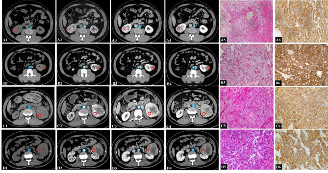
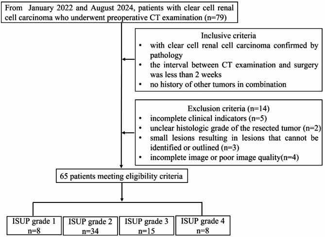
Methods: This retrospective study, approved by the institutional review board, included 65 patients (median age: 58.40 ± 10.84 years) who were diagnosed with ccRCC based on the nucleolar grading of the International Society of Urological Pathology (ISUP). All patients underwent preoperative abdominal enhanced CT between January 2022 and August 2024. CT features from the unenhanced, corticomedullary, nephrographic, and delayed phases were analyzed, and the extracellular volume fraction (ECV) of ccRCC was calculated by measuring CT values from regions of interest in both the unenhanced and nephrographic phases. Statistical significance was evaluated for differences in these parameters across the four ISUP grades. Additionally, diagnostic efficiency was assessed using receiver operating characteristic (ROC) curve analysis.
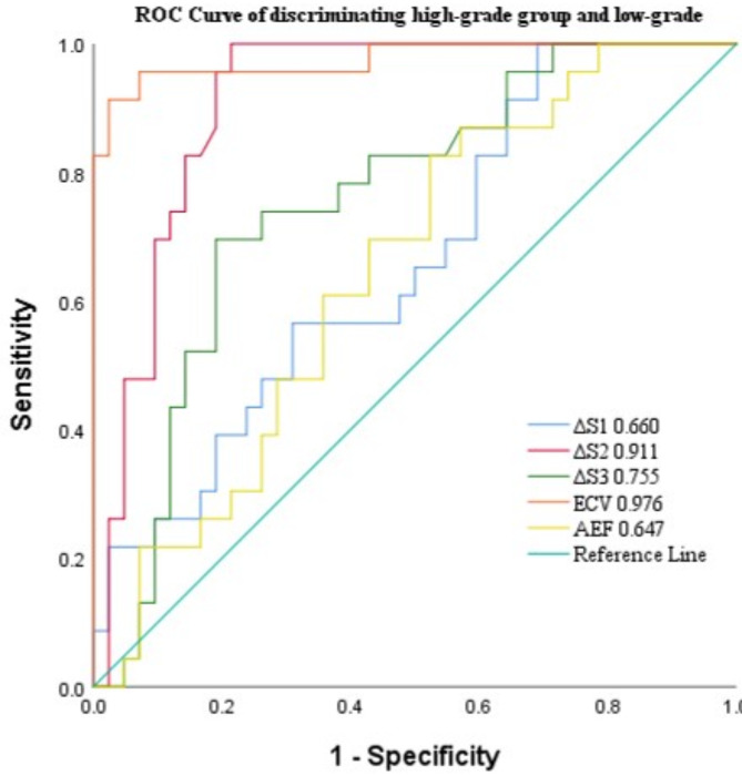
Results: The ECV showed significant differences across the four ISUP grades of ccRCC, its potential as an important predictor of high-grade ccRCC (P = 0.035). The ROC curve analysis indicated that ECV exhibited the highest diagnostic efficacy for assessing the lower- and higher- pathological grade of ccRCC, with an area under the ROC curve of 0.976. The optimal diagnostic threshold for ECV was determined to be 41.64%, with a sensitivity of 91.31% and a specificity of 97.62%.
Conclusions: ECV derived from enhanced CT has the potential to function as an in vivo biomarker for distinguishing between lower- and higher-grade ccRCC. This quantitative measure provides diagnostic value that extends beyond traditional qualitative CT features, offering a more precise and objective assessment of tumor grade.
Cancer ImagingONCOLOGY-RADIOLOGY, NUCLEAR MEDICINE & MEDICAL IMAGING
CiteScore
7.00
自引率
0.00%
发文量
66
审稿时长
>12 weeks
期刊介绍:
Cancer Imaging is an open access, peer-reviewed journal publishing original articles, reviews and editorials written by expert international radiologists working in oncology.
The journal encompasses CT, MR, PET, ultrasound, radionuclide and multimodal imaging in all kinds of malignant tumours, plus new developments, techniques and innovations. Topics of interest include:
Breast Imaging
Chest
Complications of treatment
Ear, Nose & Throat
Gastrointestinal
Hepatobiliary & Pancreatic
Imaging biomarkers
Interventional
Lymphoma
Measurement of tumour response
Molecular functional imaging
Musculoskeletal
Neuro oncology
Nuclear Medicine
Paediatric.

 求助内容:
求助内容: 应助结果提醒方式:


