Xin-Yu Liu, Zhi-Lin Yuan, Fu-Ze Cong, Li Mao, Xiu-Li Li, Zhen Zhou, Jing Ren, Yuan Li, Yan Zhang, Yong-Lan He, Hua-Dan Xue, Zheng-Yu Jin
{"title":"Deep learning assisted detection and segmentation of uterine fibroids using multi-orientation magnetic resonance imaging","authors":"Xin-Yu Liu, Zhi-Lin Yuan, Fu-Ze Cong, Li Mao, Xiu-Li Li, Zhen Zhou, Jing Ren, Yuan Li, Yan Zhang, Yong-Lan He, Hua-Dan Xue, Zheng-Yu Jin","doi":"10.1007/s00261-025-04934-8","DOIUrl":null,"url":null,"abstract":"<div><h3>Purpose</h3><p>To develop deep learning models for automated detection and segmentation of uterine fibroids using multi-orientation MRI.</p><h3>Methods</h3><p>Pre-treatment sagittal and axial T2-weighted MRI scans acquired from patients diagnosed with uterine fibroids were collected. The proposed segmentation models were constructed based on the three-dimensional nnU-Net framework. Fibroid detection efficacy was assessed, with subgroup analyses by size and location. The segmentation performance was evaluated using Dice similarity coefficients (DSCs), 95% Hausdorff distance (HD95), and average surface distance (ASD).</p><h3>Results</h3><p>The internal dataset comprised 299 patients who were divided into the training set (n = 239) and the internal test set (n = 60). The external dataset comprised 45 patients. The sagittal T2WI model and the axial T2WI model demonstrated recalls of 74.4%/76.4% and precision of 98.9%/97.9% for fibroid detection in the internal test set. The models achieved recalls of 93.7%/95.3% for fibroids ≥ 4 cm. The recalls for International Federation of Gynecology and Obstetrics (FIGO) type 2–5, FIGO types 0\\1\\2(submucous), fibroids FIGO types 5\\6\\7(subserous) were 100%/100%, 73.3%/78.6%, and 80.3%/81.9%, respectively. The proposed models demonstrated good performance in segmentation of the uterine fibroids with mean DSCs of 0.789 and 0.804, HD95s of 9.996 and 10.855 mm, and ASDs of 2.035 and 2.115 mm in the internal test set, and with mean DSCs of 0.834 and 0.818, HD95s of 9.971 and 11.874 mm, and ASDs of 2.031 and 2.273 mm in the external test set.</p><h3>Conclusion</h3><p>The proposed deep learning models showed promise as reliable methods for automating the detection and segmentation of the uterine fibroids, particularly those of clinical relevance.</p><h3>Graphical abstract</h3>\n<div><figure><div><div><picture><source><img></source></picture></div></div></figure></div></div>","PeriodicalId":7126,"journal":{"name":"Abdominal Radiology","volume":"50 10","pages":"4927 - 4938"},"PeriodicalIF":2.2000,"publicationDate":"2025-04-05","publicationTypes":"Journal Article","fieldsOfStudy":null,"isOpenAccess":false,"openAccessPdf":"","citationCount":"0","resultStr":null,"platform":"Semanticscholar","paperid":null,"PeriodicalName":"Abdominal Radiology","FirstCategoryId":"3","ListUrlMain":"https://link.springer.com/article/10.1007/s00261-025-04934-8","RegionNum":3,"RegionCategory":"医学","ArticlePicture":[],"TitleCN":null,"AbstractTextCN":null,"PMCID":null,"EPubDate":"","PubModel":"","JCR":"Q2","JCRName":"RADIOLOGY, NUCLEAR MEDICINE & MEDICAL IMAGING","Score":null,"Total":0}
引用次数: 0
Abstract
Purpose
To develop deep learning models for automated detection and segmentation of uterine fibroids using multi-orientation MRI.
Methods
Pre-treatment sagittal and axial T2-weighted MRI scans acquired from patients diagnosed with uterine fibroids were collected. The proposed segmentation models were constructed based on the three-dimensional nnU-Net framework. Fibroid detection efficacy was assessed, with subgroup analyses by size and location. The segmentation performance was evaluated using Dice similarity coefficients (DSCs), 95% Hausdorff distance (HD95), and average surface distance (ASD).
Results
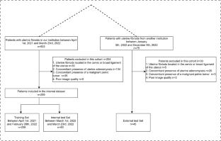
The internal dataset comprised 299 patients who were divided into the training set (n = 239) and the internal test set (n = 60). The external dataset comprised 45 patients. The sagittal T2WI model and the axial T2WI model demonstrated recalls of 74.4%/76.4% and precision of 98.9%/97.9% for fibroid detection in the internal test set. The models achieved recalls of 93.7%/95.3% for fibroids ≥ 4 cm. The recalls for International Federation of Gynecology and Obstetrics (FIGO) type 2–5, FIGO types 0\1\2(submucous), fibroids FIGO types 5\6\7(subserous) were 100%/100%, 73.3%/78.6%, and 80.3%/81.9%, respectively. The proposed models demonstrated good performance in segmentation of the uterine fibroids with mean DSCs of 0.789 and 0.804, HD95s of 9.996 and 10.855 mm, and ASDs of 2.035 and 2.115 mm in the internal test set, and with mean DSCs of 0.834 and 0.818, HD95s of 9.971 and 11.874 mm, and ASDs of 2.031 and 2.273 mm in the external test set.
Conclusion
The proposed deep learning models showed promise as reliable methods for automating the detection and segmentation of the uterine fibroids, particularly those of clinical relevance.
期刊介绍:
Abdominal Radiology seeks to meet the professional needs of the abdominal radiologist by publishing clinically pertinent original, review and practice related articles on the gastrointestinal and genitourinary tracts and abdominal interventional and radiologic procedures. Case reports are generally not accepted unless they are the first report of a new disease or condition, or part of a special solicited section.
Reasons to Publish Your Article in Abdominal Radiology:
· Official journal of the Society of Abdominal Radiology (SAR)
· Published in Cooperation with:
European Society of Gastrointestinal and Abdominal Radiology (ESGAR)
European Society of Urogenital Radiology (ESUR)
Asian Society of Abdominal Radiology (ASAR)
· Efficient handling and Expeditious review
· Author feedback is provided in a mentoring style
· Global readership
· Readers can earn CME credits


 求助内容:
求助内容: 应助结果提醒方式:


