{"title":"Clinical Presentation and Surgical Management of a Grynfelt Hernia: Report of a Clinical Case and Literature Review.","authors":"Pabel Ruben Carbajal Cabrera, Ruben Daniel Pérez López, Yunuen Ailyn Morales Tercero, Itzel Ocampo Barrero","doi":"10.1155/cris/5634242","DOIUrl":null,"url":null,"abstract":"<p><p><b>Background:</b> Grynfelt's lumbar hernia is the rarest of all abdominal wall hernias, accounting for between 1.5% and 2% of cases, with only 300-350 instances described to date. Lumbar hernias can be congenital or acquired, often triggered by trauma or surgery (iatrogenic). Diagnosis is clinical and confirmed via computed tomography. Surgical intervention is required for resolution, with repair performed either through open or laparoscopic surgery. <b>Material and Methods:</b> We present the case of a young female with no prior surgical or traumatic history, in whom the diagnosis of Grynfelt's hernia was made. <b>Results:</b> The patient underwent elective left lumbotomy surgery with hernioplasty using a supra-aponeurotic polypropylene mesh. Postsurgical recovery was adequate, and she was discharged 4 h after surgery. Follow-up in the general surgery outpatient clinic occurred at 20 days, 1, 3, and 6 months, with no recurrence, complications, or incidents. <b>Conclusion:</b> Grynfelt's hernia is a rare entity that requires a high index of suspicion for accurate diagnosis. Although cases are often asymptomatic, untreated hernias can lead to significant morbidity. Early recognition and timely surgical intervention are crucial for symptom relief and prevention of complications. In this case report, surgical management involved hernioplasty through a left lumbotomy approach, repairing the hernia defect and reducing the hernia content. Supra-aponeurotic mesh was placed to ensure adequate closure. Given the rarity of this pathology, no specific management guidelines exist in the literature. Therefore, the decision for this type of repair was based on intraoperative findings. Further research is needed to clarify management strategies and optimize outcomes for patients with Grynfelt's hernia.</p>","PeriodicalId":9600,"journal":{"name":"Case Reports in Surgery","volume":"2025 ","pages":"5634242"},"PeriodicalIF":0.5000,"publicationDate":"2025-03-27","publicationTypes":"Journal Article","fieldsOfStudy":null,"isOpenAccess":false,"openAccessPdf":"https://www.ncbi.nlm.nih.gov/pmc/articles/PMC11968149/pdf/","citationCount":"0","resultStr":null,"platform":"Semanticscholar","paperid":null,"PeriodicalName":"Case Reports in Surgery","FirstCategoryId":"1085","ListUrlMain":"https://doi.org/10.1155/cris/5634242","RegionNum":0,"RegionCategory":null,"ArticlePicture":[],"TitleCN":null,"AbstractTextCN":null,"PMCID":null,"EPubDate":"2025/1/1 0:00:00","PubModel":"eCollection","JCR":"Q4","JCRName":"SURGERY","Score":null,"Total":0}
引用次数: 0
Abstract
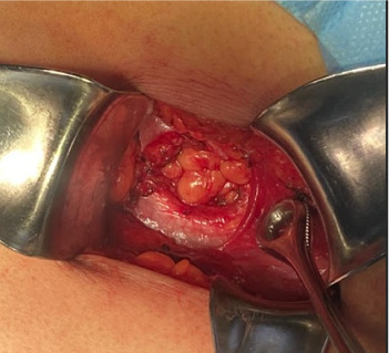
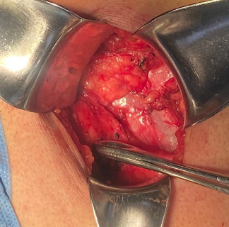
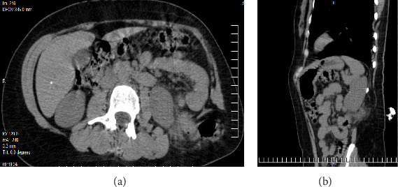
Background: Grynfelt's lumbar hernia is the rarest of all abdominal wall hernias, accounting for between 1.5% and 2% of cases, with only 300-350 instances described to date. Lumbar hernias can be congenital or acquired, often triggered by trauma or surgery (iatrogenic). Diagnosis is clinical and confirmed via computed tomography. Surgical intervention is required for resolution, with repair performed either through open or laparoscopic surgery. Material and Methods: We present the case of a young female with no prior surgical or traumatic history, in whom the diagnosis of Grynfelt's hernia was made. Results: The patient underwent elective left lumbotomy surgery with hernioplasty using a supra-aponeurotic polypropylene mesh. Postsurgical recovery was adequate, and she was discharged 4 h after surgery. Follow-up in the general surgery outpatient clinic occurred at 20 days, 1, 3, and 6 months, with no recurrence, complications, or incidents. Conclusion: Grynfelt's hernia is a rare entity that requires a high index of suspicion for accurate diagnosis. Although cases are often asymptomatic, untreated hernias can lead to significant morbidity. Early recognition and timely surgical intervention are crucial for symptom relief and prevention of complications. In this case report, surgical management involved hernioplasty through a left lumbotomy approach, repairing the hernia defect and reducing the hernia content. Supra-aponeurotic mesh was placed to ensure adequate closure. Given the rarity of this pathology, no specific management guidelines exist in the literature. Therefore, the decision for this type of repair was based on intraoperative findings. Further research is needed to clarify management strategies and optimize outcomes for patients with Grynfelt's hernia.

 求助内容:
求助内容: 应助结果提醒方式:


