{"title":"Improving the diagnostic performance of contrast-enhanced mammography through lesion conspicuity and enhancement quantification.","authors":"Iris Allajbeu, Muzna Nanaa, Roido Manavaki, Vasiliki Papalouka, Ioana Bene, Nicholas Payne, Elisabetta Giannotti, Thiemo van Nijnatten, Fleur Kilburn-Toppin, Nuala Healy, Fiona Gilbert","doi":"10.1007/s00330-025-11501-8","DOIUrl":null,"url":null,"abstract":"<p><strong>Objectives: </strong>To analyze qualitative and quantitative enhancement of breast lesions on CEM and their impact on specificity and overall diagnostic performance in predicting malignancy. A secondary objective was to compare lesion enhancement patterns between CEM and contrast-enhanced (CE)-MRI.</p><p><strong>Methods: </strong>The cohort included screening and symptomatic cases from CEM research studies (December 2016-March 2023) with an identifiable lesion. Three breast radiologists independently assessed lesion conspicuity as low, moderate, or high, based on the BI-RADS CEM lexicon. Lesion enhancement was quantified by drawing two regions of interest representing lesion and background parenchyma, to calculate contrast enhancement from the early (CE<sub>early</sub>) and late (CE<sub>late</sub>) views. Area-under-the-curve (AUC) was used to assess diagnostic performance, with thresholds determined using the maximum Youden index. Cohen's κ was used to measure agreement between CEM and DCE-MRI enhancement patterns. p-values < 0.05 were deemed statistically significant.</p><p><strong>Results: </strong>From 503 CEM studies, 143 BI-RADS 2-5 lesions were analyzed. Lesion conspicuity was significantly associated with lesion histology (p < 0.001), contrast enhancement metrics (CE<sub>early</sub>, CE<sub>late</sub>), and enhancement patterns on CEM recombined images. CE<sub>early</sub> performed better in differentiating malignant from benign lesions or background parenchymal enhancement (BPE), with AUC values of 0.83 and 0.88 and 90% specificity in distinguishing BPE from cancers. There was fair/moderate agreement between lesion enhancement patterns on CEM and DCE-MRI (Cohen's κ = 0.35, p < 0.001), with a higher agreement for lesions exhibiting a wash-out pattern (Cohen's κ = 0.5, p < 0.001).</p><p><strong>Conclusion: </strong>Both conspicuity and quantification of lesion enhancement can improve CEM specificity in predicting malignancy, with CE<sub>early</sub> offering the best diagnostic performance.</p><p><strong>Key points: </strong>Question Quantifying lesion enhancement conspicuity on contrast-enhanced mammography (CEM) has demonstrated potential in differentiating malignancy from benign lesions and BPE. Finding Contrast from the early recombined view (CEearly) performed better in discriminating malignant from benign lesions and BPE, with 90% specificity for BPE vs cancers. Clinical relevance Conspicuity and quantification of lesion enhancement on CEM can improve the specificity and overall diagnostic performance of CEM in cancer detection. Implementation of conspicuity thresholds in routine CEM interpretation could potentially reduce unnecessary recalls and benign biopsies.</p>","PeriodicalId":12076,"journal":{"name":"European Radiology","volume":" ","pages":"6385-6397"},"PeriodicalIF":4.7000,"publicationDate":"2025-10-01","publicationTypes":"Journal Article","fieldsOfStudy":null,"isOpenAccess":false,"openAccessPdf":"https://www.ncbi.nlm.nih.gov/pmc/articles/PMC12417241/pdf/","citationCount":"0","resultStr":null,"platform":"Semanticscholar","paperid":null,"PeriodicalName":"European Radiology","FirstCategoryId":"3","ListUrlMain":"https://doi.org/10.1007/s00330-025-11501-8","RegionNum":2,"RegionCategory":"医学","ArticlePicture":[],"TitleCN":null,"AbstractTextCN":null,"PMCID":null,"EPubDate":"2025/4/3 0:00:00","PubModel":"Epub","JCR":"Q1","JCRName":"RADIOLOGY, NUCLEAR MEDICINE & MEDICAL IMAGING","Score":null,"Total":0}
引用次数: 0
Abstract
Objectives: To analyze qualitative and quantitative enhancement of breast lesions on CEM and their impact on specificity and overall diagnostic performance in predicting malignancy. A secondary objective was to compare lesion enhancement patterns between CEM and contrast-enhanced (CE)-MRI.
Methods: The cohort included screening and symptomatic cases from CEM research studies (December 2016-March 2023) with an identifiable lesion. Three breast radiologists independently assessed lesion conspicuity as low, moderate, or high, based on the BI-RADS CEM lexicon. Lesion enhancement was quantified by drawing two regions of interest representing lesion and background parenchyma, to calculate contrast enhancement from the early (CEearly) and late (CElate) views. Area-under-the-curve (AUC) was used to assess diagnostic performance, with thresholds determined using the maximum Youden index. Cohen's κ was used to measure agreement between CEM and DCE-MRI enhancement patterns. p-values < 0.05 were deemed statistically significant.
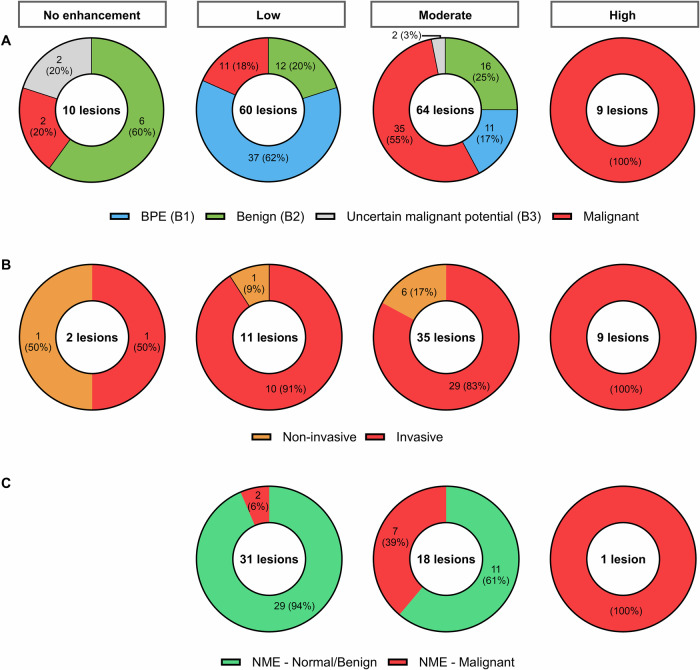
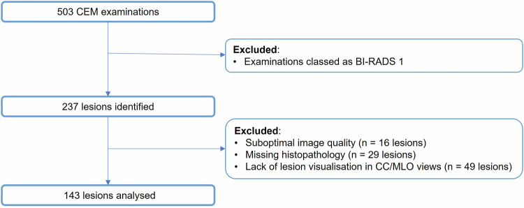
Results: From 503 CEM studies, 143 BI-RADS 2-5 lesions were analyzed. Lesion conspicuity was significantly associated with lesion histology (p < 0.001), contrast enhancement metrics (CEearly, CElate), and enhancement patterns on CEM recombined images. CEearly performed better in differentiating malignant from benign lesions or background parenchymal enhancement (BPE), with AUC values of 0.83 and 0.88 and 90% specificity in distinguishing BPE from cancers. There was fair/moderate agreement between lesion enhancement patterns on CEM and DCE-MRI (Cohen's κ = 0.35, p < 0.001), with a higher agreement for lesions exhibiting a wash-out pattern (Cohen's κ = 0.5, p < 0.001).
Conclusion: Both conspicuity and quantification of lesion enhancement can improve CEM specificity in predicting malignancy, with CEearly offering the best diagnostic performance.
Key points: Question Quantifying lesion enhancement conspicuity on contrast-enhanced mammography (CEM) has demonstrated potential in differentiating malignancy from benign lesions and BPE. Finding Contrast from the early recombined view (CEearly) performed better in discriminating malignant from benign lesions and BPE, with 90% specificity for BPE vs cancers. Clinical relevance Conspicuity and quantification of lesion enhancement on CEM can improve the specificity and overall diagnostic performance of CEM in cancer detection. Implementation of conspicuity thresholds in routine CEM interpretation could potentially reduce unnecessary recalls and benign biopsies.
期刊介绍:
European Radiology (ER) continuously updates scientific knowledge in radiology by publication of strong original articles and state-of-the-art reviews written by leading radiologists. A well balanced combination of review articles, original papers, short communications from European radiological congresses and information on society matters makes ER an indispensable source for current information in this field.
This is the Journal of the European Society of Radiology, and the official journal of a number of societies.
From 2004-2008 supplements to European Radiology were published under its companion, European Radiology Supplements, ISSN 1613-3749.

 求助内容:
求助内容: 应助结果提醒方式:


