Katharina Scheidt, Fabian Kropla, Dirk Winkler, Robert Möbius, Martin Vychopen, Johannes Wach, Erdem Güresir, Ronny Grunert
{"title":"3D-printed skull model for enhancing training in external ventricular drainage within medical education.","authors":"Katharina Scheidt, Fabian Kropla, Dirk Winkler, Robert Möbius, Martin Vychopen, Johannes Wach, Erdem Güresir, Ronny Grunert","doi":"10.1186/s41205-025-00263-0","DOIUrl":null,"url":null,"abstract":"<p><strong>Background: </strong>The importance of reducing error rates in invasive procedures has led to the development of teaching phantoms. In collaboration with surgeons and engineers at the University Hospital of Leipzig, a new 3D-printed simulation model for external ventricular drainage was created. This model includes system-relevant components such as the ventricular system, the surrounding brain tissue and the skull bone to be trephined. The methodology for developing the simulation model is described in detail. Additionally, the system was initially evaluated by neurosurgeons using a Likert scale. Future studies are planned to assess the system's accuracy and perform comparative analyses.</p><p><strong>Methods: </strong>The data required for analysis were extracted from medical images. The phantom consists of three components: the ventricular system, the brain mass, and the skull bone. The bone component was fabricated via 3D printing using a realistic hard polyamide, PA12. The ventricular system was also 3D printed as a hollow structure using a flexible material, Elastic Resin 50 A from Formlabs. The brain tissue was modeled via a cast gelatin mold. The cerebrospinal fluid was a water solution.</p><p><strong>Results: </strong>The system's initial tests successfully simulated cerebrospinal fluid flow through the tube into the ventricular system. The skull can be trepanned. Additional materials are required at the drilling sites because of chip formation. A more pointed cannula than usual can puncture the ventricular system. With a concentration of 30 g/l, gelatin is a realistic imitation of brain tissue.</p><p><strong>Conclusion: </strong>All essential components of the skull, brain and ventricle exhibit a degree of realism that has never been achieved before. In terms of its design and reproducibility, the model is exceptionally well suited for training and consolidating methods and procedures as part of a realistic training program for the placement of external ventricular drainage.</p>","PeriodicalId":72036,"journal":{"name":"3D printing in medicine","volume":"11 1","pages":"16"},"PeriodicalIF":3.1000,"publicationDate":"2025-04-03","publicationTypes":"Journal Article","fieldsOfStudy":null,"isOpenAccess":false,"openAccessPdf":"https://www.ncbi.nlm.nih.gov/pmc/articles/PMC11969789/pdf/","citationCount":"0","resultStr":null,"platform":"Semanticscholar","paperid":null,"PeriodicalName":"3D printing in medicine","FirstCategoryId":"1085","ListUrlMain":"https://doi.org/10.1186/s41205-025-00263-0","RegionNum":0,"RegionCategory":null,"ArticlePicture":[],"TitleCN":null,"AbstractTextCN":null,"PMCID":null,"EPubDate":"","PubModel":"","JCR":"Q1","JCRName":"RADIOLOGY, NUCLEAR MEDICINE & MEDICAL IMAGING","Score":null,"Total":0}
引用次数: 0
Abstract
Background: The importance of reducing error rates in invasive procedures has led to the development of teaching phantoms. In collaboration with surgeons and engineers at the University Hospital of Leipzig, a new 3D-printed simulation model for external ventricular drainage was created. This model includes system-relevant components such as the ventricular system, the surrounding brain tissue and the skull bone to be trephined. The methodology for developing the simulation model is described in detail. Additionally, the system was initially evaluated by neurosurgeons using a Likert scale. Future studies are planned to assess the system's accuracy and perform comparative analyses.
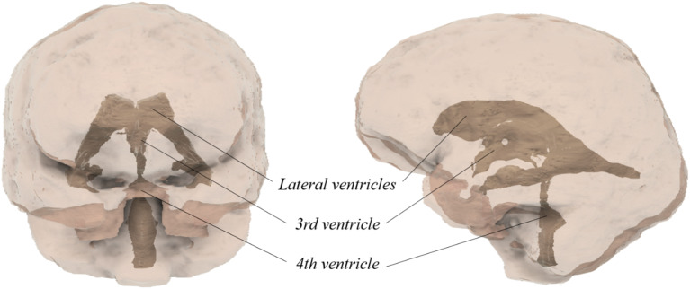
Methods: The data required for analysis were extracted from medical images. The phantom consists of three components: the ventricular system, the brain mass, and the skull bone. The bone component was fabricated via 3D printing using a realistic hard polyamide, PA12. The ventricular system was also 3D printed as a hollow structure using a flexible material, Elastic Resin 50 A from Formlabs. The brain tissue was modeled via a cast gelatin mold. The cerebrospinal fluid was a water solution.
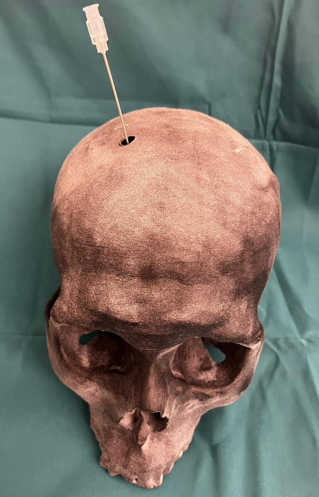
Results: The system's initial tests successfully simulated cerebrospinal fluid flow through the tube into the ventricular system. The skull can be trepanned. Additional materials are required at the drilling sites because of chip formation. A more pointed cannula than usual can puncture the ventricular system. With a concentration of 30 g/l, gelatin is a realistic imitation of brain tissue.
Conclusion: All essential components of the skull, brain and ventricle exhibit a degree of realism that has never been achieved before. In terms of its design and reproducibility, the model is exceptionally well suited for training and consolidating methods and procedures as part of a realistic training program for the placement of external ventricular drainage.

 求助内容:
求助内容: 应助结果提醒方式:


