Conrado Dias do Nascimento Neto, Laisa Kindely Ramos de Oliveira, Amy Brian Costa E Silva, Patrícia Roccon Bianchi, André Gustavo de Sousa Galdino, Daniela Nascimento Silva
{"title":"A new eggshell-derived calcium phosphate bioceramic for tissue engineering: cytotoxicity and histomorphometric study.","authors":"Conrado Dias do Nascimento Neto, Laisa Kindely Ramos de Oliveira, Amy Brian Costa E Silva, Patrícia Roccon Bianchi, André Gustavo de Sousa Galdino, Daniela Nascimento Silva","doi":"10.1590/acb402625","DOIUrl":null,"url":null,"abstract":"<p><strong>Purpose: </strong>To evaluate cytotoxicity and tissue repair of a new chicken eggshell-derived bioceramic (hydroxyapatite/dicalcium phosphate anhydrous-HA/DCPA).</p><p><strong>Methods: </strong>Cytotoxicity was evaluated in fibroblasts (L cell, L-929) by MTT test. Tissue repair of HA/DCPA was compared to HA/β-TCP bioceramic (Maxresorb-MXR). Two critical-sized bone defects (CSDs) were drilled in the calvarial of 24 Wistar rats and filled with one of the biomaterials. The animals were euthanized after 30, 60, and 90 days, and bone specimens were examined by histomorphometric analyses, scanning electron microscopy, and energy-dispersive X-ray spectroscopy. The percentages of newly formed bone, connective tissue, remaining biomaterial, and total tissue repair area were compared between groups using Student's t-test and analysis of variance (p ≤ 0.05).</p><p><strong>Results: </strong>HA/DCPA did not exhibit any cytotoxicity. CSDs contained newly formed bone from the defect margins and from ossification centers interspersed throughout the biomaterials. At 30 days, HA/DCPA group had a significantly larger total tissue repair area than MXR group (p = 0.047). No differences were observed between groups regarding variables studied (p > 0.05).</p><p><strong>Conclusion: </strong>HA/DCPA is non-cytotoxic. This cement promoted new bone formation and tissue filling of the entire defect area with degree of biomaterial degradation similar to HA/β-TCP, proving to be equally suitable and successful for bone regeneration.</p>","PeriodicalId":93850,"journal":{"name":"Acta cirurgica brasileira","volume":"40 ","pages":"e402625"},"PeriodicalIF":1.3000,"publicationDate":"2025-03-31","publicationTypes":"Journal Article","fieldsOfStudy":null,"isOpenAccess":false,"openAccessPdf":"https://www.ncbi.nlm.nih.gov/pmc/articles/PMC11960574/pdf/","citationCount":"0","resultStr":null,"platform":"Semanticscholar","paperid":null,"PeriodicalName":"Acta cirurgica brasileira","FirstCategoryId":"1085","ListUrlMain":"https://doi.org/10.1590/acb402625","RegionNum":0,"RegionCategory":null,"ArticlePicture":[],"TitleCN":null,"AbstractTextCN":null,"PMCID":null,"EPubDate":"2025/1/1 0:00:00","PubModel":"eCollection","JCR":"","JCRName":"","Score":null,"Total":0}
引用次数: 0
Abstract
Purpose: To evaluate cytotoxicity and tissue repair of a new chicken eggshell-derived bioceramic (hydroxyapatite/dicalcium phosphate anhydrous-HA/DCPA).
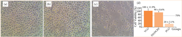
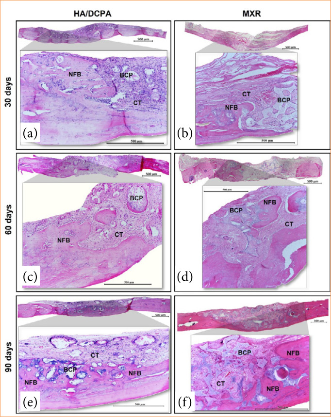
Methods: Cytotoxicity was evaluated in fibroblasts (L cell, L-929) by MTT test. Tissue repair of HA/DCPA was compared to HA/β-TCP bioceramic (Maxresorb-MXR). Two critical-sized bone defects (CSDs) were drilled in the calvarial of 24 Wistar rats and filled with one of the biomaterials. The animals were euthanized after 30, 60, and 90 days, and bone specimens were examined by histomorphometric analyses, scanning electron microscopy, and energy-dispersive X-ray spectroscopy. The percentages of newly formed bone, connective tissue, remaining biomaterial, and total tissue repair area were compared between groups using Student's t-test and analysis of variance (p ≤ 0.05).
Results: HA/DCPA did not exhibit any cytotoxicity. CSDs contained newly formed bone from the defect margins and from ossification centers interspersed throughout the biomaterials. At 30 days, HA/DCPA group had a significantly larger total tissue repair area than MXR group (p = 0.047). No differences were observed between groups regarding variables studied (p > 0.05).
Conclusion: HA/DCPA is non-cytotoxic. This cement promoted new bone formation and tissue filling of the entire defect area with degree of biomaterial degradation similar to HA/β-TCP, proving to be equally suitable and successful for bone regeneration.

 求助内容:
求助内容: 应助结果提醒方式:


