Andrea Varazzani, Pierre Petolat, Louis Brochet, Alice Prevost, Nicolas Graillon, Antoine Pierrefeu
{"title":"Disabling Plantar Keloids Treated with Brachytherapy and Free Flap Reconstruction: A Case Report.","authors":"Andrea Varazzani, Pierre Petolat, Louis Brochet, Alice Prevost, Nicolas Graillon, Antoine Pierrefeu","doi":"10.1159/000545053","DOIUrl":null,"url":null,"abstract":"<p><strong>Introduction: </strong>Keloids result from a disorganized fibroproliferative collagen response that extends beyond the original wound margins and fails to regress. They are associated with a high recurrence rate despite various treatment options. Keloids on the sole of the foot are exceedingly rare and pose significant challenges for both patients and surgeons. To date, only 15 cases of plantar keloids have been described in the English literature. Management options for this region include observation with custom-made footwear, steroid injections, surgery alone, or surgery followed by multimodal therapy. This report presents the first documented case of a plantar keloid treated with surgical excision, brachytherapy, and free flap reconstruction.</p><p><strong>Case presentation: </strong>We describe the case of a plantar keloid treated with surgical excision followed by high-dose-rate brachytherapy and free flap reconstruction. At 18 months postoperatively, the patient was ambulating independently without crutches, though hypersensitivity persisted. The scars at the donor site and the medial ankle exhibited keloids, but all scars subjected to brachytherapy were normal.</p><p><strong>Conclusion: </strong>The free flap approach may not be the optimal reconstruction method for plantar keloids, as skin grafts appear to have better outcomes, according to the literature. Also, in our patient, brachytherapy has proven its effectiveness in preventing further keloid formation as shown in the literature.</p>","PeriodicalId":9619,"journal":{"name":"Case Reports in Dermatology","volume":"17 1","pages":"96-105"},"PeriodicalIF":0.8000,"publicationDate":"2025-03-11","publicationTypes":"Journal Article","fieldsOfStudy":null,"isOpenAccess":false,"openAccessPdf":"https://www.ncbi.nlm.nih.gov/pmc/articles/PMC11961157/pdf/","citationCount":"0","resultStr":null,"platform":"Semanticscholar","paperid":null,"PeriodicalName":"Case Reports in Dermatology","FirstCategoryId":"1085","ListUrlMain":"https://doi.org/10.1159/000545053","RegionNum":0,"RegionCategory":null,"ArticlePicture":[],"TitleCN":null,"AbstractTextCN":null,"PMCID":null,"EPubDate":"2025/1/1 0:00:00","PubModel":"eCollection","JCR":"Q4","JCRName":"DERMATOLOGY","Score":null,"Total":0}
引用次数: 0
Abstract
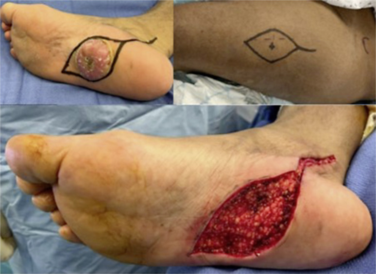
Introduction: Keloids result from a disorganized fibroproliferative collagen response that extends beyond the original wound margins and fails to regress. They are associated with a high recurrence rate despite various treatment options. Keloids on the sole of the foot are exceedingly rare and pose significant challenges for both patients and surgeons. To date, only 15 cases of plantar keloids have been described in the English literature. Management options for this region include observation with custom-made footwear, steroid injections, surgery alone, or surgery followed by multimodal therapy. This report presents the first documented case of a plantar keloid treated with surgical excision, brachytherapy, and free flap reconstruction.
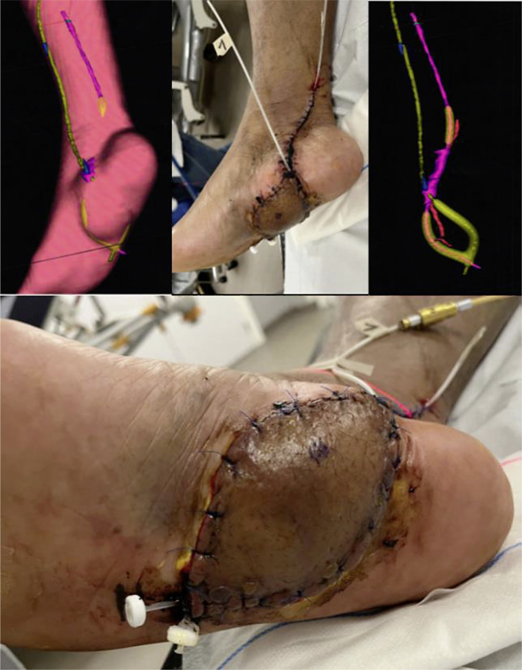
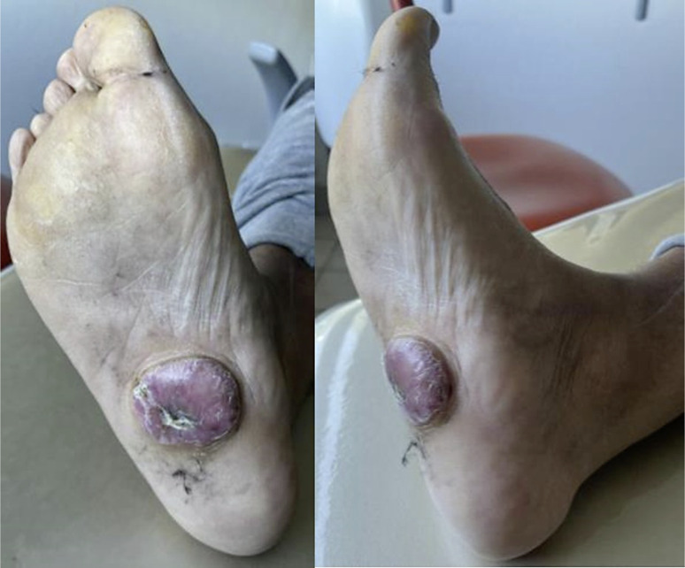
Case presentation: We describe the case of a plantar keloid treated with surgical excision followed by high-dose-rate brachytherapy and free flap reconstruction. At 18 months postoperatively, the patient was ambulating independently without crutches, though hypersensitivity persisted. The scars at the donor site and the medial ankle exhibited keloids, but all scars subjected to brachytherapy were normal.
Conclusion: The free flap approach may not be the optimal reconstruction method for plantar keloids, as skin grafts appear to have better outcomes, according to the literature. Also, in our patient, brachytherapy has proven its effectiveness in preventing further keloid formation as shown in the literature.

 求助内容:
求助内容: 应助结果提醒方式:


