Analysis of the best time-point for 18F-FDG PET/CT delayed imaging in patients of small colorectal cancer liver metastasis with hypothyroidism based on diagnostic efficacy and image standardized uptake values
IF 2.5 4区 医学Q2 RADIOLOGY, NUCLEAR MEDICINE & MEDICAL IMAGING
{"title":"Analysis of the best time-point for 18F-FDG PET/CT delayed imaging in patients of small colorectal cancer liver metastasis with hypothyroidism based on diagnostic efficacy and image standardized uptake values","authors":"Yusong Pei, Yanan Tong, Zhiguo Wang, Xinxin Qiao, Yanqing Liu, Guoxu Zhang","doi":"10.1007/s12149-025-02045-4","DOIUrl":null,"url":null,"abstract":"<div><h3>Objective</h3><p>This study analyzes the role of positron emission tomography/computed tomography (PET/CT) in the diagnosis of small (< 10 mm) colorectal cancer liver metastasis (CRLM) lesions in patients with hypothyroidism. In particular, the impact of the best time for delayed imaging on improving diagnostic efficacy.</p><h3>Methods</h3><p>We retrospectively analyzed 231 patients with small CRLM lesions with hypothyroidism who underwent dual time-point 18F-FDG PET/CT imaging. Based on the previous studies and clinical practice experience, 120–190 min was selected as the time range for delayed imaging, divided into eight teams in 10-min groups. The delayed images of the eight time periods were first analyzed and compared for diagnostic efficacy, and second analyzed and compared for standardized uptake value (SUV) and of PET/CT images to observe the trend of SUV values over time.</p><h3>Results</h3><p>The results of diagnostic efficacy analysis indicated that the 180-min delay group had the highest diagnostic efficacy (sensitivity, specificity, and accuracy). Comparison of the SUV values with the delay time analysis showed that maximum standardized uptake value (SUVmax) increased with the delay time, and the normal liver tissue (SUVmean) decreased with the delay time, which resulted in the gradual increase in the ratio of the lesion to the normal liver tissue (TNR). By selecting the time-point with the highest TNR ratio and stable SUV value, and combining the results of diagnostic efficacy, this study successfully verified the best imaging time-point. After comprehensive consideration, 180 min was determined as the best imaging time-point, when the TNR reached the highest, the SUV value was stable, and the diagnostic efficacy was best.</p><h3>Conclusions</h3><p>In this study, the impact of delayed imaging on the diagnostic efficacy and SUV value of PET/CT images in patients of small CRLM with hypothyroidism was shown intuitively, and the changing pattern of SUV at different time points was also observed. The best time-point for PET/CT delayed imaging was determined to be 180 min, which provides a new scanning program for the diagnosis in patients of small CRLM with hypothyroidism.</p></div>","PeriodicalId":8007,"journal":{"name":"Annals of Nuclear Medicine","volume":"39 7","pages":"707 - 715"},"PeriodicalIF":2.5000,"publicationDate":"2025-04-02","publicationTypes":"Journal Article","fieldsOfStudy":null,"isOpenAccess":false,"openAccessPdf":"","citationCount":"0","resultStr":null,"platform":"Semanticscholar","paperid":null,"PeriodicalName":"Annals of Nuclear Medicine","FirstCategoryId":"3","ListUrlMain":"https://link.springer.com/article/10.1007/s12149-025-02045-4","RegionNum":4,"RegionCategory":"医学","ArticlePicture":[],"TitleCN":null,"AbstractTextCN":null,"PMCID":null,"EPubDate":"","PubModel":"","JCR":"Q2","JCRName":"RADIOLOGY, NUCLEAR MEDICINE & MEDICAL IMAGING","Score":null,"Total":0}
引用次数: 0
Abstract
Objective
This study analyzes the role of positron emission tomography/computed tomography (PET/CT) in the diagnosis of small (< 10 mm) colorectal cancer liver metastasis (CRLM) lesions in patients with hypothyroidism. In particular, the impact of the best time for delayed imaging on improving diagnostic efficacy.
Methods
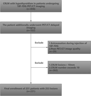
We retrospectively analyzed 231 patients with small CRLM lesions with hypothyroidism who underwent dual time-point 18F-FDG PET/CT imaging. Based on the previous studies and clinical practice experience, 120–190 min was selected as the time range for delayed imaging, divided into eight teams in 10-min groups. The delayed images of the eight time periods were first analyzed and compared for diagnostic efficacy, and second analyzed and compared for standardized uptake value (SUV) and of PET/CT images to observe the trend of SUV values over time.
Results
The results of diagnostic efficacy analysis indicated that the 180-min delay group had the highest diagnostic efficacy (sensitivity, specificity, and accuracy). Comparison of the SUV values with the delay time analysis showed that maximum standardized uptake value (SUVmax) increased with the delay time, and the normal liver tissue (SUVmean) decreased with the delay time, which resulted in the gradual increase in the ratio of the lesion to the normal liver tissue (TNR). By selecting the time-point with the highest TNR ratio and stable SUV value, and combining the results of diagnostic efficacy, this study successfully verified the best imaging time-point. After comprehensive consideration, 180 min was determined as the best imaging time-point, when the TNR reached the highest, the SUV value was stable, and the diagnostic efficacy was best.
Conclusions
In this study, the impact of delayed imaging on the diagnostic efficacy and SUV value of PET/CT images in patients of small CRLM with hypothyroidism was shown intuitively, and the changing pattern of SUV at different time points was also observed. The best time-point for PET/CT delayed imaging was determined to be 180 min, which provides a new scanning program for the diagnosis in patients of small CRLM with hypothyroidism.
期刊介绍:
Annals of Nuclear Medicine is an official journal of the Japanese Society of Nuclear Medicine. It develops the appropriate application of radioactive substances and stable nuclides in the field of medicine.
The journal promotes the exchange of ideas and information and research in nuclear medicine and includes the medical application of radionuclides and related subjects. It presents original articles, short communications, reviews and letters to the editor.

 求助内容:
求助内容: 应助结果提醒方式:


