Nikoline D Frølich, Jeannette D Andersen, Helle D Zacho
{"title":"The frequency and characterization of ovarian metastasis from nonovarian cancers using 18F-fluorodeoxyglucose PET/CT.","authors":"Nikoline D Frølich, Jeannette D Andersen, Helle D Zacho","doi":"10.1093/bjro/tzaf004","DOIUrl":null,"url":null,"abstract":"<p><strong>Objective: </strong>Assessing the frequency of ovarian metastasis from nonovarian cancer (N-OC) and evaluate whether any PET-derived parameters can distinguish metastasis from primary ovarian cancer.</p><p><strong>Methods: </strong>Patients undergoing FDG PET/CT due to suspected ovarian malignancy from 2006 to 2021 with subsequent histologically proven ovarian metastasis from N-OC were included. Exclusion criteria included ovarian metastasis diagnosed prior to PET/CT or >3 months after. Baseline characteristics were collected from electronic medical records, and PET/CT data were analysed using Siemens syngo.via software.</p><p><strong>Results: </strong>Patients (<i>N</i> =1502) were scanned for suspected ovarian malignancies. Sixty-five patients (4%) were included. The most common origin of metastases was lower gastrointestinal cancer (<i>n</i> = 29, 45%), followed by gynaecological cancer (<i>n</i> = 10, 15%) and breast cancer (<i>n</i> = 9, 14%). Among patients with previous cancer history (<i>n</i> = 26), 18 experienced ovarian metastases from a known cancer. Time from primary diagnosis to ovarian metastasis ranged from 47 days to 11.4 years. There were no differences in maximized standardized uptake value, peak standardized uptake value, or clinical parameters between ovarian metastases and primary ovarian tumours.</p><p><strong>Conclusion: </strong>The frequency of ovarian metastases from N-OCs was 4%, the most common origin of metastases was lower gastrointestinal tract. Previous cancer history is an important factor in assessing an unknown tumour of the ovary, as metastases can occur several years later. No PET or clinical parameters were useful for separating primary ovarian cancer from ovarian metastases.</p><p><strong>Advances in knowledge: </strong>The study finds a low frequency of ovarian metastasis from N-OC and indicates that no PET or clinical parameters can distinguish ovarian metastasis from primary ovarian cancer.</p>","PeriodicalId":72419,"journal":{"name":"BJR open","volume":"7 1","pages":"tzaf004"},"PeriodicalIF":2.1000,"publicationDate":"2025-03-18","publicationTypes":"Journal Article","fieldsOfStudy":null,"isOpenAccess":false,"openAccessPdf":"https://www.ncbi.nlm.nih.gov/pmc/articles/PMC11954554/pdf/","citationCount":"0","resultStr":null,"platform":"Semanticscholar","paperid":null,"PeriodicalName":"BJR open","FirstCategoryId":"1085","ListUrlMain":"https://doi.org/10.1093/bjro/tzaf004","RegionNum":0,"RegionCategory":null,"ArticlePicture":[],"TitleCN":null,"AbstractTextCN":null,"PMCID":null,"EPubDate":"2025/1/1 0:00:00","PubModel":"eCollection","JCR":"","JCRName":"","Score":null,"Total":0}
引用次数: 0
Abstract
Objective: Assessing the frequency of ovarian metastasis from nonovarian cancer (N-OC) and evaluate whether any PET-derived parameters can distinguish metastasis from primary ovarian cancer.
Methods: Patients undergoing FDG PET/CT due to suspected ovarian malignancy from 2006 to 2021 with subsequent histologically proven ovarian metastasis from N-OC were included. Exclusion criteria included ovarian metastasis diagnosed prior to PET/CT or >3 months after. Baseline characteristics were collected from electronic medical records, and PET/CT data were analysed using Siemens syngo.via software.
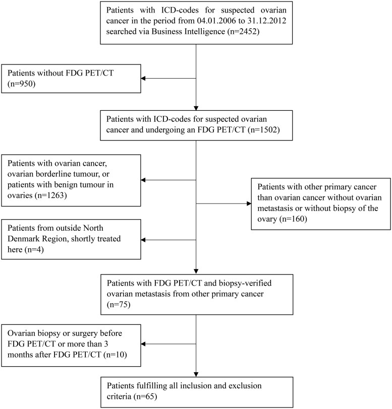
Results: Patients (N =1502) were scanned for suspected ovarian malignancies. Sixty-five patients (4%) were included. The most common origin of metastases was lower gastrointestinal cancer (n = 29, 45%), followed by gynaecological cancer (n = 10, 15%) and breast cancer (n = 9, 14%). Among patients with previous cancer history (n = 26), 18 experienced ovarian metastases from a known cancer. Time from primary diagnosis to ovarian metastasis ranged from 47 days to 11.4 years. There were no differences in maximized standardized uptake value, peak standardized uptake value, or clinical parameters between ovarian metastases and primary ovarian tumours.
Conclusion: The frequency of ovarian metastases from N-OCs was 4%, the most common origin of metastases was lower gastrointestinal tract. Previous cancer history is an important factor in assessing an unknown tumour of the ovary, as metastases can occur several years later. No PET or clinical parameters were useful for separating primary ovarian cancer from ovarian metastases.
Advances in knowledge: The study finds a low frequency of ovarian metastasis from N-OC and indicates that no PET or clinical parameters can distinguish ovarian metastasis from primary ovarian cancer.

 求助内容:
求助内容: 应助结果提醒方式:


