Julio César Moreno-Alfonso, Sara Hernández Martín, Lidia Ayuso González, Alberto Perez Martínez
{"title":"[Abdominal pain and inconclusive ultrasound: a CT scan may not always be the best next step]","authors":"Julio César Moreno-Alfonso, Sara Hernández Martín, Lidia Ayuso González, Alberto Perez Martínez","doi":"10.31053/1853.0605.v82.n1.44691","DOIUrl":null,"url":null,"abstract":"<p><p>A 4-year-old male presented to the emergency department with fever, mucous diarrhea, abdominal pain, and vomiting for 36 hours. On abdominal palpation, he had generalized pain without rebound and increased hydroaereal sounds. The blood count showed leukocytosis and elevated C-reactive protein, and an ultrasound revealed intestinal loops with abundant content, increased peristalsis, and a normal appendix. Due to the suspicion of invasive gastroenteritis, he was admitted for observation. Due to the persistence of symptoms, a new ultrasound was performed which showed pelvic free fluid but did not identify the appendix. An abdominal radiography was previously performed, which showed an image compatible with appendicolith. With the suspicion of appendicitis, surgery was indicated, and an appendicular peritonitis was identified. The patient recovered well and was discharged seven days later. Diagnosing appendicitis in children can be a complex process. The most sensitive diagnostic tests are ultrasound and tomography. Abdominal radiography, however, is a widely available test with low radiation and has a positive predictive value of 90% for appendicitis (detecting appendicolith or periappendicular 'air silence'). In ambiguous cases, an abdominal X-ray could avoid invasive tests that require sedation or high doses of radiation.</p>","PeriodicalId":38814,"journal":{"name":"Revista de la Facultad de Ciencias Medicas de Cordoba","volume":"82 1","pages":"206-214"},"PeriodicalIF":0.0000,"publicationDate":"2025-03-31","publicationTypes":"Journal Article","fieldsOfStudy":null,"isOpenAccess":false,"openAccessPdf":"https://www.ncbi.nlm.nih.gov/pmc/articles/PMC12057710/pdf/","citationCount":"0","resultStr":null,"platform":"Semanticscholar","paperid":null,"PeriodicalName":"Revista de la Facultad de Ciencias Medicas de Cordoba","FirstCategoryId":"1085","ListUrlMain":"https://doi.org/10.31053/1853.0605.v82.n1.44691","RegionNum":0,"RegionCategory":null,"ArticlePicture":[],"TitleCN":null,"AbstractTextCN":null,"PMCID":null,"EPubDate":"","PubModel":"","JCR":"Q3","JCRName":"Medicine","Score":null,"Total":0}
引用次数: 0
Abstract
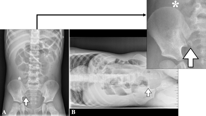
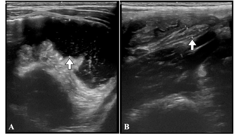
A 4-year-old male presented to the emergency department with fever, mucous diarrhea, abdominal pain, and vomiting for 36 hours. On abdominal palpation, he had generalized pain without rebound and increased hydroaereal sounds. The blood count showed leukocytosis and elevated C-reactive protein, and an ultrasound revealed intestinal loops with abundant content, increased peristalsis, and a normal appendix. Due to the suspicion of invasive gastroenteritis, he was admitted for observation. Due to the persistence of symptoms, a new ultrasound was performed which showed pelvic free fluid but did not identify the appendix. An abdominal radiography was previously performed, which showed an image compatible with appendicolith. With the suspicion of appendicitis, surgery was indicated, and an appendicular peritonitis was identified. The patient recovered well and was discharged seven days later. Diagnosing appendicitis in children can be a complex process. The most sensitive diagnostic tests are ultrasound and tomography. Abdominal radiography, however, is a widely available test with low radiation and has a positive predictive value of 90% for appendicitis (detecting appendicolith or periappendicular 'air silence'). In ambiguous cases, an abdominal X-ray could avoid invasive tests that require sedation or high doses of radiation.
期刊介绍:
The Journal of the Faculty of Medical Sciences is a scientific publication of the Secretariat of Science and Technology of the Faculty of Medical Sciences of the National University of Cordoba. Its objective is to disseminate and promote research work related to Medical and Biological Sciences. It publishes scientific works of national and international professionals on different topics related to health sciences from the field of medicine, nursing, kinesiology, diagnostic imaging, phonoaudiology, nutrition, public health, chemical sciences, dentistry and related.

 求助内容:
求助内容: 应助结果提醒方式:


