Dual-channel pulse-dye densitometry can enable correction of fluorescent targeted and control agent plasma input function differences for quantitative paired-agent molecular imaging: a simulation study.
Cody C Rounds, Yichen Feng, Sanjana Pannem, Jovan Brankov, Kimberly S Samkoe, Kenneth M Tichauer
{"title":"Dual-channel pulse-dye densitometry can enable correction of fluorescent targeted and control agent plasma input function differences for quantitative paired-agent molecular imaging: a simulation study.","authors":"Cody C Rounds, Yichen Feng, Sanjana Pannem, Jovan Brankov, Kimberly S Samkoe, Kenneth M Tichauer","doi":"10.1117/1.JBO.30.4.046001","DOIUrl":null,"url":null,"abstract":"<p><strong>Significance: </strong>Paired-agent fluorescent molecular imaging approaches involve co-administration of a control (untargeted) imaging agent with a molecularly targeted agent to account for non-specific effects and quantify binding potential (BP)-a parameter proportional to the concentration of the targeted biomolecule. Accurate BP estimation often requires correction for differences in targeted and control agent plasma input functions (PIFs).</p><p><strong>Aim: </strong>We provide a simulation-based evaluation of whether dual-channel pulse dye densitometry (PDD) can be used to measure the PIFs of co-administered targeted and control imaging agents, to enable accurate BP estimation.</p><p><strong>Approach: </strong>Monte-Carlo simulations of light propagation were carried out using the anatomy and optical properties of a finger, as well as experimentally measured PIFs of co-administered anti-epidermal growth factor receptor fluorescent affibody, ABY-029, and IRDye 680LT, a control imaging agent from past mouse experiments. The accuracy of PIF shape estimation from PDD and PIF difference correction was evaluated by assessing BP estimation accuracy in a simulated \"tumor\" tissue.</p><p><strong>Results: </strong>\"Tumor\" BP measurements using deconvolution correction with noise-free PIFs versus PDD-measured PIFs were compared. The relative error in PDD PIF deconvolution BP estimation was <math><mrow><mn>2</mn> <mo>±</mo> <mn>1</mn> <mo>%</mo></mrow> </math> . No statistical difference was found between the estimated BP via deconvolution correction with true PIFs and the estimated BP via the reconstructed PIFs using the proposed PAF-PDD methodology.</p><p><strong>Conclusions: </strong>These results highlight the potential for developing a PDD instrument that can directly measure targeted and control agent PIFs and be used to correct for any PIF differences between agents for more quantitative estimates of BP in paired-agent imaging studies.</p>","PeriodicalId":15264,"journal":{"name":"Journal of Biomedical Optics","volume":"30 4","pages":"046001"},"PeriodicalIF":2.9000,"publicationDate":"2025-04-01","publicationTypes":"Journal Article","fieldsOfStudy":null,"isOpenAccess":false,"openAccessPdf":"https://www.ncbi.nlm.nih.gov/pmc/articles/PMC11954597/pdf/","citationCount":"0","resultStr":null,"platform":"Semanticscholar","paperid":null,"PeriodicalName":"Journal of Biomedical Optics","FirstCategoryId":"3","ListUrlMain":"https://doi.org/10.1117/1.JBO.30.4.046001","RegionNum":3,"RegionCategory":"医学","ArticlePicture":[],"TitleCN":null,"AbstractTextCN":null,"PMCID":null,"EPubDate":"2025/3/29 0:00:00","PubModel":"Epub","JCR":"Q2","JCRName":"BIOCHEMICAL RESEARCH METHODS","Score":null,"Total":0}
引用次数: 0
Abstract
Significance: Paired-agent fluorescent molecular imaging approaches involve co-administration of a control (untargeted) imaging agent with a molecularly targeted agent to account for non-specific effects and quantify binding potential (BP)-a parameter proportional to the concentration of the targeted biomolecule. Accurate BP estimation often requires correction for differences in targeted and control agent plasma input functions (PIFs).
Aim: We provide a simulation-based evaluation of whether dual-channel pulse dye densitometry (PDD) can be used to measure the PIFs of co-administered targeted and control imaging agents, to enable accurate BP estimation.
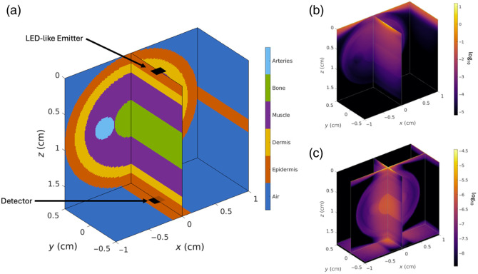
Approach: Monte-Carlo simulations of light propagation were carried out using the anatomy and optical properties of a finger, as well as experimentally measured PIFs of co-administered anti-epidermal growth factor receptor fluorescent affibody, ABY-029, and IRDye 680LT, a control imaging agent from past mouse experiments. The accuracy of PIF shape estimation from PDD and PIF difference correction was evaluated by assessing BP estimation accuracy in a simulated "tumor" tissue.
Results: "Tumor" BP measurements using deconvolution correction with noise-free PIFs versus PDD-measured PIFs were compared. The relative error in PDD PIF deconvolution BP estimation was . No statistical difference was found between the estimated BP via deconvolution correction with true PIFs and the estimated BP via the reconstructed PIFs using the proposed PAF-PDD methodology.
Conclusions: These results highlight the potential for developing a PDD instrument that can directly measure targeted and control agent PIFs and be used to correct for any PIF differences between agents for more quantitative estimates of BP in paired-agent imaging studies.
期刊介绍:
The Journal of Biomedical Optics publishes peer-reviewed papers on the use of modern optical technology for improved health care and biomedical research.

 求助内容:
求助内容: 应助结果提醒方式:


