Davide Burrascano, Barbara Verro, Gaetano Ottoveggio, Ada Maria Florena, Carmelo Saraniti
{"title":"Laryngeal Frame Involvement as The First Sign of Wegener's Granulomatosis.","authors":"Davide Burrascano, Barbara Verro, Gaetano Ottoveggio, Ada Maria Florena, Carmelo Saraniti","doi":"10.22038/ijorl.2024.81617.3746","DOIUrl":null,"url":null,"abstract":"<p><strong>Introduction: </strong>Granulomatosis with Polyangiitis (GPA), also known as Wegener's Granulomatosis, is an ANCA-associated vasculitis that primarily affects small vessels, leading to necrotizing granulomatous reactions in the airways and small vessels. The etiology remains uncertain.</p><p><strong>Case report: </strong>We report the case of a woman in her 70s, who was previously tracheostomized at another facility and was presented to our attention with glottic-subglottic stenosis. We performed a lysis of glottic synechia and subglottic debulking via transoral laser microsurgery, yielding satisfactory results over the short term. However, a relapse occurred within two months, along with ulcerative lesions on the nasal septum. Biopsies revealed multinucleated giant cells and inflammation suggestive of vasculitis. Based on the histological and clinical features, a diagnosis of vasculitis was considered. Anti-Neutrophil Cytoplasmic Antibodies testing was positive. A rheumatological examination confirmed the hypothesis of Granulomatosis with Polyangiitis. The lack of typical symptoms was the main reason for the delayed diagnosis.</p><p><strong>Conclusion: </strong>Involvement of the subglottic region and the upper portion of the trachea is a rare but severe complication of GPA. The current literature reports only few cases of laryngeal stenosis, with poor prognosis. Histological examinations of biopsied laryngeal tissue showed significant but non-specific inflammation, contributing to the delay in diagnosis. There are still no precise guidelines for the surgical treatment of subglottic stenosis. This case underscores the importance of considering laryngeal involvement in GPA for early diagnosis and timely intervention to prevent serious complications in order to improve patient outcomes.</p>","PeriodicalId":14607,"journal":{"name":"Iranian Journal of Otorhinolaryngology","volume":"37 2","pages":"95-98"},"PeriodicalIF":0.0000,"publicationDate":"2025-01-01","publicationTypes":"Journal Article","fieldsOfStudy":null,"isOpenAccess":false,"openAccessPdf":"https://www.ncbi.nlm.nih.gov/pmc/articles/PMC11949434/pdf/","citationCount":"0","resultStr":null,"platform":"Semanticscholar","paperid":null,"PeriodicalName":"Iranian Journal of Otorhinolaryngology","FirstCategoryId":"1085","ListUrlMain":"https://doi.org/10.22038/ijorl.2024.81617.3746","RegionNum":0,"RegionCategory":null,"ArticlePicture":[],"TitleCN":null,"AbstractTextCN":null,"PMCID":null,"EPubDate":"","PubModel":"","JCR":"Q3","JCRName":"Medicine","Score":null,"Total":0}
引用次数: 0
Abstract
Introduction: Granulomatosis with Polyangiitis (GPA), also known as Wegener's Granulomatosis, is an ANCA-associated vasculitis that primarily affects small vessels, leading to necrotizing granulomatous reactions in the airways and small vessels. The etiology remains uncertain.
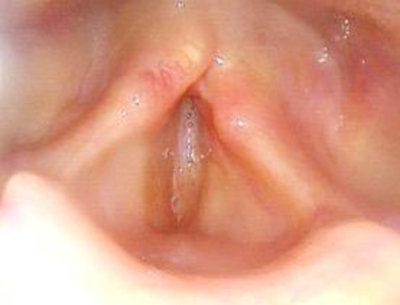
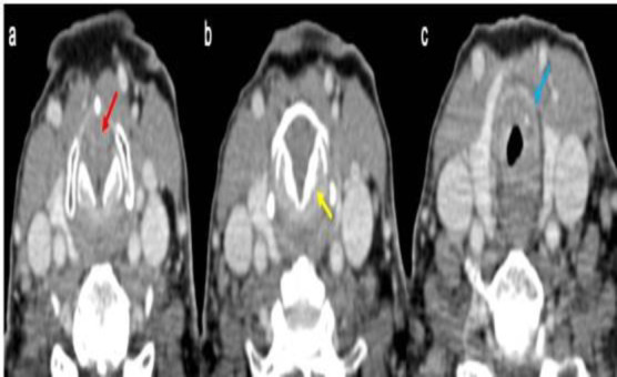
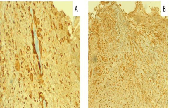
Case report: We report the case of a woman in her 70s, who was previously tracheostomized at another facility and was presented to our attention with glottic-subglottic stenosis. We performed a lysis of glottic synechia and subglottic debulking via transoral laser microsurgery, yielding satisfactory results over the short term. However, a relapse occurred within two months, along with ulcerative lesions on the nasal septum. Biopsies revealed multinucleated giant cells and inflammation suggestive of vasculitis. Based on the histological and clinical features, a diagnosis of vasculitis was considered. Anti-Neutrophil Cytoplasmic Antibodies testing was positive. A rheumatological examination confirmed the hypothesis of Granulomatosis with Polyangiitis. The lack of typical symptoms was the main reason for the delayed diagnosis.
Conclusion: Involvement of the subglottic region and the upper portion of the trachea is a rare but severe complication of GPA. The current literature reports only few cases of laryngeal stenosis, with poor prognosis. Histological examinations of biopsied laryngeal tissue showed significant but non-specific inflammation, contributing to the delay in diagnosis. There are still no precise guidelines for the surgical treatment of subglottic stenosis. This case underscores the importance of considering laryngeal involvement in GPA for early diagnosis and timely intervention to prevent serious complications in order to improve patient outcomes.
多血管炎肉芽肿病(Granulomatosis with Polyangiitis, GPA),又称Wegener肉芽肿病,是一种anca相关的血管炎,主要影响小血管,导致气道和小血管坏死性肉芽肿反应。病因尚不清楚。病例报告:我们报告一位70多岁的女性,她之前在另一家医院做过气管造口术,并因声门-声门下狭窄而引起我们的注意。我们通过经口激光显微手术进行了声门粘连的溶解和声门下的减压,在短期内取得了令人满意的结果。然而,在两个月内复发,并伴有鼻中隔溃疡性病变。活检显示多核巨细胞和提示血管炎的炎症。根据组织学和临床特征,考虑血管炎的诊断。抗中性粒细胞胞浆抗体试验阳性。风湿病检查证实了肉芽肿合并多血管炎的假设。缺乏典型症状是延误诊断的主要原因。结论:累及声门下区及气管上部是GPA罕见但严重的并发症。目前文献报道的喉部狭窄病例很少,预后较差。喉部活检组织的组织学检查显示明显但非特异性炎症,导致诊断延迟。对于声门下狭窄的手术治疗仍没有精确的指南。本病例强调了在GPA中考虑喉受累的重要性,以便早期诊断和及时干预,以防止严重并发症,以改善患者的预后。

 求助内容:
求助内容: 应助结果提醒方式:


