Yao Zhou, Wenying Chen, Shiyu Feng, Shuangchao Liu, Cheng Chen, Bingxu Yao, Xiao Li Shen
{"title":"Ochratoxin A-induced mitochondrial pathway apoptosis and ferroptosis by promoting glycolysis","authors":"Yao Zhou, Wenying Chen, Shiyu Feng, Shuangchao Liu, Cheng Chen, Bingxu Yao, Xiao Li Shen","doi":"10.1007/s10495-025-02109-w","DOIUrl":null,"url":null,"abstract":"<div><p>Ochratoxin A (OTA), a toxic secondary metabolite recognized for its harmful effects on the kidneys, and it is commonly present in various foods and animal feeds. Although there have been few reports on the involvement of metabolic enzymes in OTA-induced nephrotoxicity and metabolic reprogramming in OTA-induced digestive tract toxicity, it remains unclear whether OTA’s primary nephrotoxic effects are mediated through metabolic reprogramming. In this study, we examined the effects of OTA and/or 2-deoxy-D-glucose (2-DG) on cell viability, levels of reactive oxygen species (ROS), glutathione (GSH), malondialdehyde (MDA), and lactic acid (LA), as well as protein levels in human proximal tubule epithelial (HK-2) cells. The results indicate that OTA leads to a reduction in GSH levels and the protein levels of Lon protease 1 (Lonp1), tumor necrosis factor receptor-associated protein 1 (TRAP1), mitochondrial pyruvate carrier 1 (MPC1), glutathione peroxidase 4 (GPX4), B-cell lymphoma-2 (Bcl-2), and Bcl-2-like protein 1 (Bcl-xl), while increasing ROS, MDA, and LA levels, as well as the protein levels of glucose transporter type 1 (GLUT1), hexokinase 2 (HK2), pyruvate kinase 2 (PKM2), ATP-dependent 6-phosphofructokinase, platelet type (PFKP), long-chain fatty acid-CoA ligase 4 (ACSL4), Bcl-2-associated X protein (Bax), and cyclophilin D (CYPD) (<i>P</i> < 0.05). In conclusion, OTA induces mitochondrial pathway apoptosis and ferroptosis by disturbing mitochondrial homeostasis via the inhibition of Lonp1 and TRAP1, thereby reducing GSH levels, increasing ROS, MDA, and LA levels, and promoting glycolysis in vitro. This is the first report on OTA-induced mitochondrial pathway apoptosis and ferroptosis facilitated by mitochondrial homeostasis imbalance-mediated glycolysis in HK-2 cells.</p></div>","PeriodicalId":8062,"journal":{"name":"Apoptosis","volume":"30 5-6","pages":"1440 - 1452"},"PeriodicalIF":8.1000,"publicationDate":"2025-04-01","publicationTypes":"Journal Article","fieldsOfStudy":null,"isOpenAccess":false,"openAccessPdf":"","citationCount":"0","resultStr":null,"platform":"Semanticscholar","paperid":null,"PeriodicalName":"Apoptosis","FirstCategoryId":"99","ListUrlMain":"https://link.springer.com/article/10.1007/s10495-025-02109-w","RegionNum":2,"RegionCategory":"生物学","ArticlePicture":[],"TitleCN":null,"AbstractTextCN":null,"PMCID":null,"EPubDate":"","PubModel":"","JCR":"Q1","JCRName":"BIOCHEMISTRY & MOLECULAR BIOLOGY","Score":null,"Total":0}
引用次数: 0
Abstract
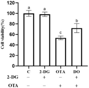
Ochratoxin A (OTA), a toxic secondary metabolite recognized for its harmful effects on the kidneys, and it is commonly present in various foods and animal feeds. Although there have been few reports on the involvement of metabolic enzymes in OTA-induced nephrotoxicity and metabolic reprogramming in OTA-induced digestive tract toxicity, it remains unclear whether OTA’s primary nephrotoxic effects are mediated through metabolic reprogramming. In this study, we examined the effects of OTA and/or 2-deoxy-D-glucose (2-DG) on cell viability, levels of reactive oxygen species (ROS), glutathione (GSH), malondialdehyde (MDA), and lactic acid (LA), as well as protein levels in human proximal tubule epithelial (HK-2) cells. The results indicate that OTA leads to a reduction in GSH levels and the protein levels of Lon protease 1 (Lonp1), tumor necrosis factor receptor-associated protein 1 (TRAP1), mitochondrial pyruvate carrier 1 (MPC1), glutathione peroxidase 4 (GPX4), B-cell lymphoma-2 (Bcl-2), and Bcl-2-like protein 1 (Bcl-xl), while increasing ROS, MDA, and LA levels, as well as the protein levels of glucose transporter type 1 (GLUT1), hexokinase 2 (HK2), pyruvate kinase 2 (PKM2), ATP-dependent 6-phosphofructokinase, platelet type (PFKP), long-chain fatty acid-CoA ligase 4 (ACSL4), Bcl-2-associated X protein (Bax), and cyclophilin D (CYPD) (P < 0.05). In conclusion, OTA induces mitochondrial pathway apoptosis and ferroptosis by disturbing mitochondrial homeostasis via the inhibition of Lonp1 and TRAP1, thereby reducing GSH levels, increasing ROS, MDA, and LA levels, and promoting glycolysis in vitro. This is the first report on OTA-induced mitochondrial pathway apoptosis and ferroptosis facilitated by mitochondrial homeostasis imbalance-mediated glycolysis in HK-2 cells.
期刊介绍:
Apoptosis, a monthly international peer-reviewed journal, focuses on the rapid publication of innovative investigations into programmed cell death. The journal aims to stimulate research on the mechanisms and role of apoptosis in various human diseases, such as cancer, autoimmune disease, viral infection, AIDS, cardiovascular disease, neurodegenerative disorders, osteoporosis, and aging. The Editor-In-Chief acknowledges the importance of advancing clinical therapies for apoptosis-related diseases. Apoptosis considers Original Articles, Reviews, Short Communications, Letters to the Editor, and Book Reviews for publication.

 求助内容:
求助内容: 应助结果提醒方式:


