Development and validation of a nomogram for predicting bone marrow involvement in lymphoma patients based on 18F-FDG PET radiomics and clinical factors
IF 2.5 4区 医学Q2 RADIOLOGY, NUCLEAR MEDICINE & MEDICAL IMAGING
{"title":"Development and validation of a nomogram for predicting bone marrow involvement in lymphoma patients based on 18F-FDG PET radiomics and clinical factors","authors":"Denglu Lu, Xinyu Zhu, Xingyu Mu, Xiaoqi Huang, Feng Wei, Lilan Qin, Qixin Liu, Wei Fu, Yanyun Deng","doi":"10.1007/s12149-025-02041-8","DOIUrl":null,"url":null,"abstract":"<div><h3>Objective</h3><p>This study aimed to develop and validate a nomogram combining <sup>18</sup>F-FDG PET radiomics and clinical factors to non-invasively predict bone marrow involvement (BMI) in patients with lymphoma.</p><h3>Methods</h3><p>A radiomics nomogram was developed using monocentric data, randomly divided into a training set (70%) and a test set (30%). Bone marrow biopsy (BMB) served as the gold standard for BMI diagnosis. Independent clinical risk factors were identified through univariate and multivariate logistic regression analyses to construct a clinical model. Radiomics features were extracted from PET and CT images and selected using least absolute shrinkage and selection operator (LASSO) regression, yielding a radiomics score (Rad<sub>score</sub>) for each patient. Models based on clinical factors, CT Rad<sub>score</sub>, and PET Rad<sub>score</sub> were established and evaluated using eight machine learning algorithms to identify the optimal prediction model. A combined model was constructed and presented as a nomogram. Model performance was assessed using the area under the receiver operating characteristic curve (AUC), calibration curves, and decision curve analysis (DCA).</p><h3>Results</h3><p>A total of 160 patients were included, of whom 70 had BMI based on BMB results. The training group comprised 112 patients (BMI: 56, without BMI: 56), while the test group included 48 patients (BMI: 14, without BMI: 34). Independent risk factors, including the number of extranodal involvements and B symptoms, were incorporated into the clinical model. In the clinical model, CT Rad<sub>score</sub>, and PET Rad<sub>score</sub>, the AUCs in the test set were 0.820 (95% CI: 0.705–0.935), 0.538 (95% CI: 0.351–0.723), and 0.836 (95% CI: 0.686–0.986). Due to the limited diagnostic performance of CT Rad<sub>score</sub>, the nomogram was constructed using PET Rad<sub>score</sub> and the clinical model. The radiomics nomogram achieved AUCs of 0.916 (95% CI: 0.865–0.967) in the training set and 0.863 (95% CI: 0.763–0.964) in the test set. Calibration curves and DCA confirmed the nomogram’s discrimination, calibration, and clinical utility in both sets.</p><h3>Conclusion</h3><p>By integrating PET Rad<sub>score</sub>, the number of extranodal involvements, and B symptoms, this <sup>18</sup>F-FDG PET radiomics-based nomogram offers a non-invasive method to predict bone marrow status in lymphoma patients, providing nuclear medicine physicians with valuable decision support for pre-treatment evaluation.</p></div>","PeriodicalId":8007,"journal":{"name":"Annals of Nuclear Medicine","volume":"39 7","pages":"663 - 675"},"PeriodicalIF":2.5000,"publicationDate":"2025-03-29","publicationTypes":"Journal Article","fieldsOfStudy":null,"isOpenAccess":false,"openAccessPdf":"","citationCount":"0","resultStr":null,"platform":"Semanticscholar","paperid":null,"PeriodicalName":"Annals of Nuclear Medicine","FirstCategoryId":"3","ListUrlMain":"https://link.springer.com/article/10.1007/s12149-025-02041-8","RegionNum":4,"RegionCategory":"医学","ArticlePicture":[],"TitleCN":null,"AbstractTextCN":null,"PMCID":null,"EPubDate":"","PubModel":"","JCR":"Q2","JCRName":"RADIOLOGY, NUCLEAR MEDICINE & MEDICAL IMAGING","Score":null,"Total":0}
引用次数: 0
Abstract
Objective
This study aimed to develop and validate a nomogram combining 18F-FDG PET radiomics and clinical factors to non-invasively predict bone marrow involvement (BMI) in patients with lymphoma.
Methods
A radiomics nomogram was developed using monocentric data, randomly divided into a training set (70%) and a test set (30%). Bone marrow biopsy (BMB) served as the gold standard for BMI diagnosis. Independent clinical risk factors were identified through univariate and multivariate logistic regression analyses to construct a clinical model. Radiomics features were extracted from PET and CT images and selected using least absolute shrinkage and selection operator (LASSO) regression, yielding a radiomics score (Radscore) for each patient. Models based on clinical factors, CT Radscore, and PET Radscore were established and evaluated using eight machine learning algorithms to identify the optimal prediction model. A combined model was constructed and presented as a nomogram. Model performance was assessed using the area under the receiver operating characteristic curve (AUC), calibration curves, and decision curve analysis (DCA).
Results
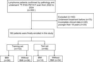
A total of 160 patients were included, of whom 70 had BMI based on BMB results. The training group comprised 112 patients (BMI: 56, without BMI: 56), while the test group included 48 patients (BMI: 14, without BMI: 34). Independent risk factors, including the number of extranodal involvements and B symptoms, were incorporated into the clinical model. In the clinical model, CT Radscore, and PET Radscore, the AUCs in the test set were 0.820 (95% CI: 0.705–0.935), 0.538 (95% CI: 0.351–0.723), and 0.836 (95% CI: 0.686–0.986). Due to the limited diagnostic performance of CT Radscore, the nomogram was constructed using PET Radscore and the clinical model. The radiomics nomogram achieved AUCs of 0.916 (95% CI: 0.865–0.967) in the training set and 0.863 (95% CI: 0.763–0.964) in the test set. Calibration curves and DCA confirmed the nomogram’s discrimination, calibration, and clinical utility in both sets.
Conclusion
By integrating PET Radscore, the number of extranodal involvements, and B symptoms, this 18F-FDG PET radiomics-based nomogram offers a non-invasive method to predict bone marrow status in lymphoma patients, providing nuclear medicine physicians with valuable decision support for pre-treatment evaluation.
期刊介绍:
Annals of Nuclear Medicine is an official journal of the Japanese Society of Nuclear Medicine. It develops the appropriate application of radioactive substances and stable nuclides in the field of medicine.
The journal promotes the exchange of ideas and information and research in nuclear medicine and includes the medical application of radionuclides and related subjects. It presents original articles, short communications, reviews and letters to the editor.

 求助内容:
求助内容: 应助结果提醒方式:


