Wounsuk Rhee, Sung Cheol Park, Hyoungmin Kim, Bong-Soon Chang, Sam Yeol Chang
{"title":"Deep learning-based prediction of cervical canal stenosis from mid-sagittal T2-weighted MRI.","authors":"Wounsuk Rhee, Sung Cheol Park, Hyoungmin Kim, Bong-Soon Chang, Sam Yeol Chang","doi":"10.1007/s00256-025-04917-2","DOIUrl":null,"url":null,"abstract":"<p><strong>Objective: </strong>This study aims to establish a large degenerative cervical myelopathy cohort and develop deep learning models for predicting cervical canal stenosis from sagittal T2-weighted MRI.</p><p><strong>Materials and methods: </strong>Data was collected retrospectively from patients who underwent a cervical spine MRI from January 2007 to December 2022 at a single institution. Ground truth labels for cervical canal stenosis were obtained from sagittal T2-weighted MRI using Kang's grade, a four-level scoring system that classifies stenosis with the degree of subarachnoid space obliteration and cord indentation. ResNet50, VGG16, MobileNetV3, and EfficientNetV2 were trained using threefold cross-validation, and the models exhibiting the largest area under the receiver operating characteristic curve (AUC) were selected to produce the ensemble model. Gradient-weighted class activation mapping was adopted for qualitative assessment. Models that incorporate demographic features were trained, and their corresponding AUCs on the test set were evaluated.</p><p><strong>Results: </strong>Of 8676 patients, 7645 were eligible for developing deep learning models, where 6880 (mean age, 56.0 ± 14.3 years, 3480 men) were used for training while 765 (mean age, 56.5 ± 14.4 years, 386 men) were set aside for testing. The ensemble model exhibited the largest AUC of 0.95 (0.94-0.97). Accuracy was 0.875 (0.851-0.898), sensitivity was 0.885 (0.855-0.915), and specificity was 0.861 (0.824-0.898). Qualitative analyses demonstrated that the models accurately pinpoint radiologic findings suggestive of cervical canal stenosis and myelopathy. Incorporation of demographic features did not result in a gain of AUC.</p><p><strong>Conclusion: </strong>We have developed deep learning models from a large degenerative cervical myelopathy cohort and thoroughly explored their robustness and explainability.</p>","PeriodicalId":21783,"journal":{"name":"Skeletal Radiology","volume":" ","pages":"2067-2076"},"PeriodicalIF":2.2000,"publicationDate":"2025-10-01","publicationTypes":"Journal Article","fieldsOfStudy":null,"isOpenAccess":false,"openAccessPdf":"https://www.ncbi.nlm.nih.gov/pmc/articles/PMC12361303/pdf/","citationCount":"0","resultStr":null,"platform":"Semanticscholar","paperid":null,"PeriodicalName":"Skeletal Radiology","FirstCategoryId":"3","ListUrlMain":"https://doi.org/10.1007/s00256-025-04917-2","RegionNum":3,"RegionCategory":"医学","ArticlePicture":[],"TitleCN":null,"AbstractTextCN":null,"PMCID":null,"EPubDate":"2025/3/28 0:00:00","PubModel":"Epub","JCR":"Q2","JCRName":"ORTHOPEDICS","Score":null,"Total":0}
引用次数: 0
Abstract
Objective: This study aims to establish a large degenerative cervical myelopathy cohort and develop deep learning models for predicting cervical canal stenosis from sagittal T2-weighted MRI.
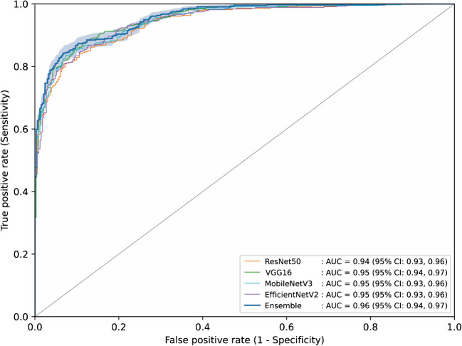
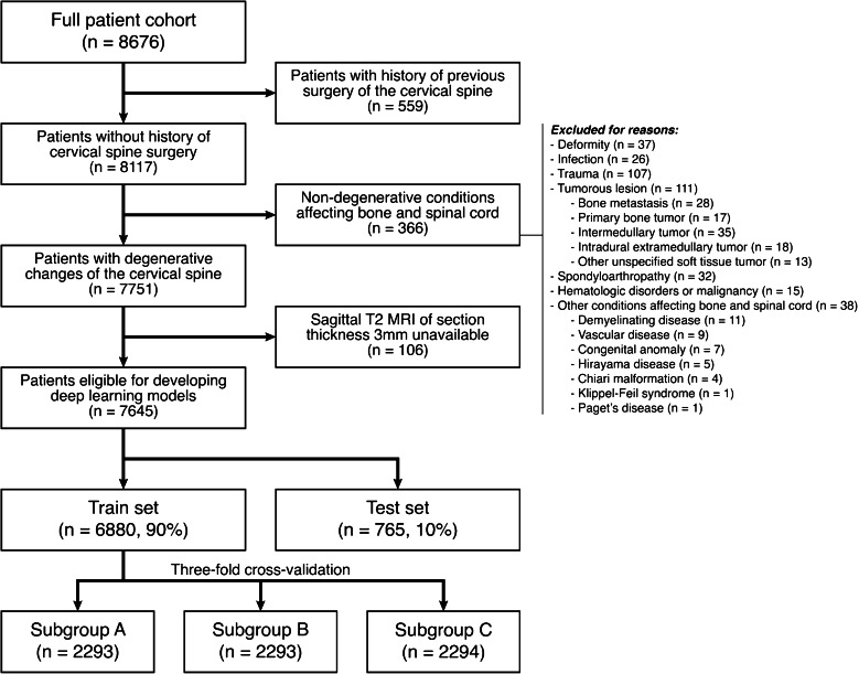
Materials and methods: Data was collected retrospectively from patients who underwent a cervical spine MRI from January 2007 to December 2022 at a single institution. Ground truth labels for cervical canal stenosis were obtained from sagittal T2-weighted MRI using Kang's grade, a four-level scoring system that classifies stenosis with the degree of subarachnoid space obliteration and cord indentation. ResNet50, VGG16, MobileNetV3, and EfficientNetV2 were trained using threefold cross-validation, and the models exhibiting the largest area under the receiver operating characteristic curve (AUC) were selected to produce the ensemble model. Gradient-weighted class activation mapping was adopted for qualitative assessment. Models that incorporate demographic features were trained, and their corresponding AUCs on the test set were evaluated.
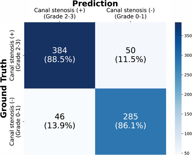
Results: Of 8676 patients, 7645 were eligible for developing deep learning models, where 6880 (mean age, 56.0 ± 14.3 years, 3480 men) were used for training while 765 (mean age, 56.5 ± 14.4 years, 386 men) were set aside for testing. The ensemble model exhibited the largest AUC of 0.95 (0.94-0.97). Accuracy was 0.875 (0.851-0.898), sensitivity was 0.885 (0.855-0.915), and specificity was 0.861 (0.824-0.898). Qualitative analyses demonstrated that the models accurately pinpoint radiologic findings suggestive of cervical canal stenosis and myelopathy. Incorporation of demographic features did not result in a gain of AUC.
Conclusion: We have developed deep learning models from a large degenerative cervical myelopathy cohort and thoroughly explored their robustness and explainability.
期刊介绍:
Skeletal Radiology provides a forum for the dissemination of current knowledge and information dealing with disorders of the musculoskeletal system including the spine. While emphasizing the radiological aspects of the many varied skeletal abnormalities, the journal also adopts an interdisciplinary approach, reflecting the membership of the International Skeletal Society. Thus, the anatomical, pathological, physiological, clinical, metabolic and epidemiological aspects of the many entities affecting the skeleton receive appropriate consideration.
This is the Journal of the International Skeletal Society and the Official Journal of the Society of Skeletal Radiology and the Australasian Musculoskelelal Imaging Group.

 求助内容:
求助内容: 应助结果提醒方式:


