Agnieszka Szmigielska, Piotr Skrzypczyk, Michał Szyszka, Magdalena Bukowska, Malwina Wojtas, Aleksandra Jakimów-Kostrzewa
{"title":"IVCA in a Boy with Multilocular Renal Cyst as a Risk Factor for Deep Vein Thrombosis.","authors":"Agnieszka Szmigielska, Piotr Skrzypczyk, Michał Szyszka, Magdalena Bukowska, Malwina Wojtas, Aleksandra Jakimów-Kostrzewa","doi":"10.34763/jmotherandchild.20242801.d-24-00045","DOIUrl":null,"url":null,"abstract":"<p><strong>Introduction: </strong>The triad of symptoms: renal defects, congenital inferior vena cava agenesis (IVCA) and deep vein thrombosis of the lower limbs make up the KILT syndrome (kidney and IVC abnormalities with leg thrombosis).</p><p><strong>Case report: </strong>A 17-year-old boy complained of periodic abdominal pain. Abdominal ultrasonography revealed a multilocular cyst in the right kidney. Physical examination showed no abnormalities, and his blood pressure was 120/80mmHg. Abdominal ultrasonography showed a cyst measuring 36×30×25mm in the right kidney hilum. Computed tomography did not show the hepatic and suprarenal sections of the inferior vena cava. Numerous varicose-dilated collateral vessels, including renal venous vessels, were found in the right kidney hilum. The collateral vessels in the tomography matched the described in the ultrasound renal cyst. MRI confirmed IVCA with no other additional vascular abnormalities. Due to the risk of deep vein thrombosis of the lower limbs, non-pharmacological antithrombotic prophylaxis was recommended.</p><p><strong>Conclusions: </strong>Early detection of inferior vena cava agenesis allows for the reduction of the risk of dangerous thrombotic complications.</p>","PeriodicalId":73842,"journal":{"name":"Journal of mother and child","volume":"28 1","pages":"146-150"},"PeriodicalIF":0.0000,"publicationDate":"2025-03-25","publicationTypes":"Journal Article","fieldsOfStudy":null,"isOpenAccess":false,"openAccessPdf":"https://www.ncbi.nlm.nih.gov/pmc/articles/PMC12002566/pdf/","citationCount":"0","resultStr":null,"platform":"Semanticscholar","paperid":null,"PeriodicalName":"Journal of mother and child","FirstCategoryId":"1085","ListUrlMain":"https://doi.org/10.34763/jmotherandchild.20242801.d-24-00045","RegionNum":0,"RegionCategory":null,"ArticlePicture":[],"TitleCN":null,"AbstractTextCN":null,"PMCID":null,"EPubDate":"2024/2/1 0:00:00","PubModel":"eCollection","JCR":"","JCRName":"","Score":null,"Total":0}
引用次数: 0
Abstract
Introduction: The triad of symptoms: renal defects, congenital inferior vena cava agenesis (IVCA) and deep vein thrombosis of the lower limbs make up the KILT syndrome (kidney and IVC abnormalities with leg thrombosis).
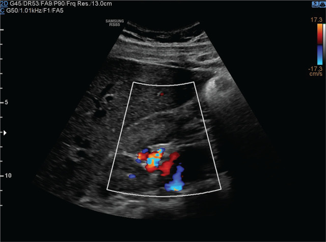
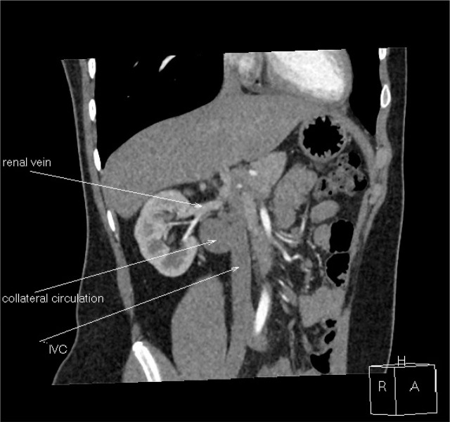
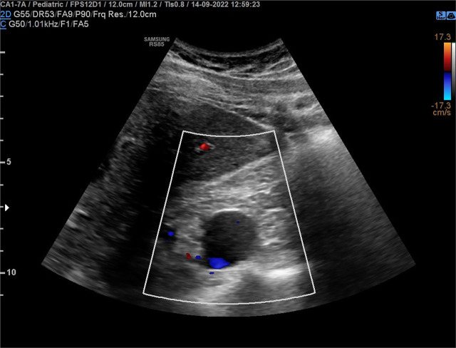
Case report: A 17-year-old boy complained of periodic abdominal pain. Abdominal ultrasonography revealed a multilocular cyst in the right kidney. Physical examination showed no abnormalities, and his blood pressure was 120/80mmHg. Abdominal ultrasonography showed a cyst measuring 36×30×25mm in the right kidney hilum. Computed tomography did not show the hepatic and suprarenal sections of the inferior vena cava. Numerous varicose-dilated collateral vessels, including renal venous vessels, were found in the right kidney hilum. The collateral vessels in the tomography matched the described in the ultrasound renal cyst. MRI confirmed IVCA with no other additional vascular abnormalities. Due to the risk of deep vein thrombosis of the lower limbs, non-pharmacological antithrombotic prophylaxis was recommended.
Conclusions: Early detection of inferior vena cava agenesis allows for the reduction of the risk of dangerous thrombotic complications.

 求助内容:
求助内容: 应助结果提醒方式:


