Dmitrii S Maltsev, Alexey N Kulikov, Alina A Kazak
{"title":"Iris Microcirculation After Selective Laser Trabeculoplasty: A Pilot Optical Coherence Tomography Angiography Study.","authors":"Dmitrii S Maltsev, Alexey N Kulikov, Alina A Kazak","doi":"10.3390/vision9010021","DOIUrl":null,"url":null,"abstract":"<p><strong>Background: </strong>This research was conducted to study changes in iris microcirculation using optical coherence tomography angiography (OCTA) in patients with primary open-angle glaucoma after selective laser trabeculoplasty (SLT).</p><p><strong>Methods: </strong>All patients received standard SLT. OCTA examination of the iris was performed before SLT and one day and seven days after SLT using RTVue-XR with a 3 mm scan pattern and follow-up function. Iris vascularity was calculated with ImageJ software (version 1.53k) as vessel density on binarized images. Correlation between absolute or percentage changes in iris vessel density and intraocular pressure (IOP) changes was calculated.</p><p><strong>Results: </strong>A total of 31 eyes (31 patients, 10 females, 70.7 ± 8.9 years) were included. Iris vessel density increased statistically significantly (<i>p</i> = 0.002) the day after SLT followed by a decrease to baseline level at one week. A statistically significant correlation (r = 0.57, <i>p</i> = 0.002) was found between the percentage change in iris vessel density the day after the procedure and IOP change at three months.</p><p><strong>Conclusion: </strong>SLT is associated with a transitory increase in iris vessel density, which can be observed with OCTA the day after the procedure. Substantial increase in iris vascularity is associated with a poorer IOP-lowering effect of SLT in eyes with open-angle glaucoma.</p>","PeriodicalId":36586,"journal":{"name":"Vision (Switzerland)","volume":"9 1","pages":""},"PeriodicalIF":1.8000,"publicationDate":"2025-03-05","publicationTypes":"Journal Article","fieldsOfStudy":null,"isOpenAccess":false,"openAccessPdf":"https://www.ncbi.nlm.nih.gov/pmc/articles/PMC11946727/pdf/","citationCount":"0","resultStr":null,"platform":"Semanticscholar","paperid":null,"PeriodicalName":"Vision (Switzerland)","FirstCategoryId":"1085","ListUrlMain":"https://doi.org/10.3390/vision9010021","RegionNum":0,"RegionCategory":null,"ArticlePicture":[],"TitleCN":null,"AbstractTextCN":null,"PMCID":null,"EPubDate":"","PubModel":"","JCR":"Q2","JCRName":"Medicine","Score":null,"Total":0}
引用次数: 0
Abstract
Background: This research was conducted to study changes in iris microcirculation using optical coherence tomography angiography (OCTA) in patients with primary open-angle glaucoma after selective laser trabeculoplasty (SLT).
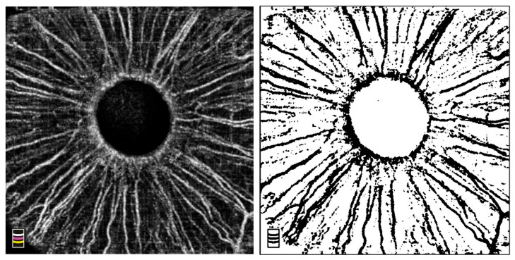
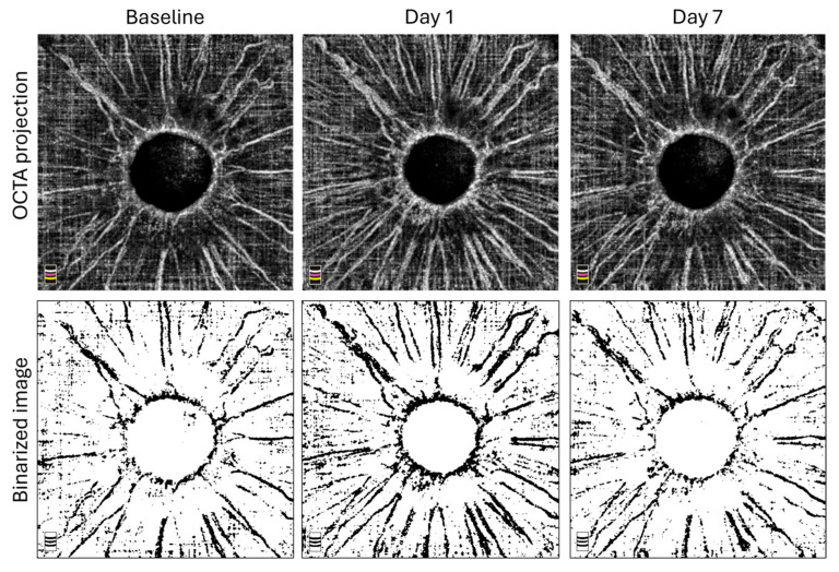
Methods: All patients received standard SLT. OCTA examination of the iris was performed before SLT and one day and seven days after SLT using RTVue-XR with a 3 mm scan pattern and follow-up function. Iris vascularity was calculated with ImageJ software (version 1.53k) as vessel density on binarized images. Correlation between absolute or percentage changes in iris vessel density and intraocular pressure (IOP) changes was calculated.
Results: A total of 31 eyes (31 patients, 10 females, 70.7 ± 8.9 years) were included. Iris vessel density increased statistically significantly (p = 0.002) the day after SLT followed by a decrease to baseline level at one week. A statistically significant correlation (r = 0.57, p = 0.002) was found between the percentage change in iris vessel density the day after the procedure and IOP change at three months.
Conclusion: SLT is associated with a transitory increase in iris vessel density, which can be observed with OCTA the day after the procedure. Substantial increase in iris vascularity is associated with a poorer IOP-lowering effect of SLT in eyes with open-angle glaucoma.

 求助内容:
求助内容: 应助结果提醒方式:


