Alicia Lim, Wei-Shan Tsai, Sridevi Thottarath, Sarega Gurudas, Taffeta Ching Ning Yamaguchi, Elizabeth Pearce, Sobha Sivaprasad
{"title":"Characterizing the Preferred Retinal Locus and Fixation Stability in Diabetic Macular Ischemia: A One-Year Study.","authors":"Alicia Lim, Wei-Shan Tsai, Sridevi Thottarath, Sarega Gurudas, Taffeta Ching Ning Yamaguchi, Elizabeth Pearce, Sobha Sivaprasad","doi":"10.3390/vision9010020","DOIUrl":null,"url":null,"abstract":"<p><p>Eyes with maculopathy usually have poor fixation stability (FS) and develop a new preferred retinal locus (PRL). The exact FS and PRL have never been studied in diabetic macular ischemia (DMI). In this one-year observational study, we recruited 79 patients (145 eyes) with evidence of DMI on optical coherence tomography angiography (OCTA). Microperimetry (MP) was performed at baseline and 52 weeks. Overall, DMI eyes demonstrated relatively stable FS without evolving into eccentric fixation over one year. When comparing the better-seeing eye (BSE) with the worse-seeing eye (WSE) in eyes with bilateral DMI, the latter presented with a larger bivariate contour ellipse area (BCEA) initially but gradually aligned with the one in the BSE at the end of the study. Conversely, the foveolar retinal sensitivity (RS) worsened significantly alongside the extension of disorganization of the retinal inner layers (DRIL) in the WSE at one year despite the best-corrected visual acuity (BCVA) being maintained. This suggests that foveolar RS might reflect the start of DMI deterioration more sensitively than BCVA.</p>","PeriodicalId":36586,"journal":{"name":"Vision (Switzerland)","volume":"9 1","pages":""},"PeriodicalIF":1.8000,"publicationDate":"2025-03-05","publicationTypes":"Journal Article","fieldsOfStudy":null,"isOpenAccess":false,"openAccessPdf":"https://www.ncbi.nlm.nih.gov/pmc/articles/PMC11945794/pdf/","citationCount":"0","resultStr":null,"platform":"Semanticscholar","paperid":null,"PeriodicalName":"Vision (Switzerland)","FirstCategoryId":"1085","ListUrlMain":"https://doi.org/10.3390/vision9010020","RegionNum":0,"RegionCategory":null,"ArticlePicture":[],"TitleCN":null,"AbstractTextCN":null,"PMCID":null,"EPubDate":"","PubModel":"","JCR":"Q2","JCRName":"Medicine","Score":null,"Total":0}
引用次数: 0
Abstract
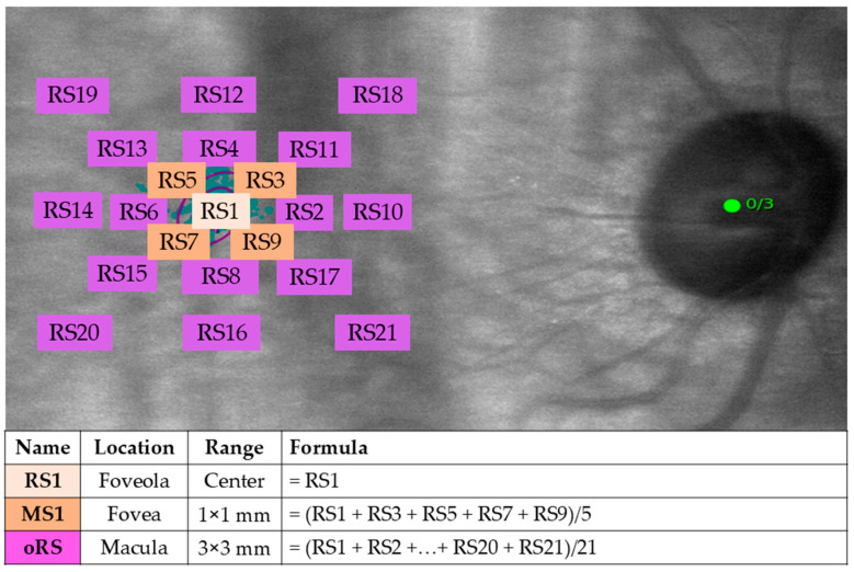
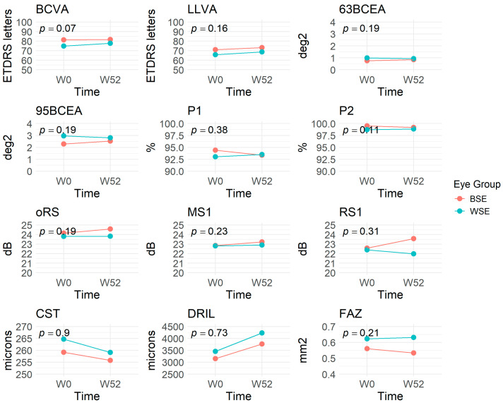
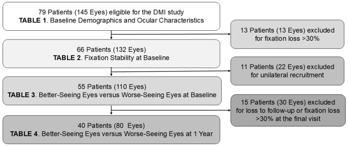
Eyes with maculopathy usually have poor fixation stability (FS) and develop a new preferred retinal locus (PRL). The exact FS and PRL have never been studied in diabetic macular ischemia (DMI). In this one-year observational study, we recruited 79 patients (145 eyes) with evidence of DMI on optical coherence tomography angiography (OCTA). Microperimetry (MP) was performed at baseline and 52 weeks. Overall, DMI eyes demonstrated relatively stable FS without evolving into eccentric fixation over one year. When comparing the better-seeing eye (BSE) with the worse-seeing eye (WSE) in eyes with bilateral DMI, the latter presented with a larger bivariate contour ellipse area (BCEA) initially but gradually aligned with the one in the BSE at the end of the study. Conversely, the foveolar retinal sensitivity (RS) worsened significantly alongside the extension of disorganization of the retinal inner layers (DRIL) in the WSE at one year despite the best-corrected visual acuity (BCVA) being maintained. This suggests that foveolar RS might reflect the start of DMI deterioration more sensitively than BCVA.

 求助内容:
求助内容: 应助结果提醒方式:


