So Hyun Park, Moon Hyung Choi, Bohyun Kim, Hyun-Soo Lee, Sungjin Yoon, Young Joon Lee, Dominik Nickel, Thomas Benkert
{"title":"Deep Learning-Accelerated Non-Contrast Abbreviated Liver MRI for Detecting Malignant Focal Hepatic Lesions: Dual-Center Validation.","authors":"So Hyun Park, Moon Hyung Choi, Bohyun Kim, Hyun-Soo Lee, Sungjin Yoon, Young Joon Lee, Dominik Nickel, Thomas Benkert","doi":"10.3348/kjr.2024.0862","DOIUrl":null,"url":null,"abstract":"<p><strong>Objective: </strong>To compare a deep learning (DL)-accelerated non-enhanced abbreviated MRI (AMRI<sub>DL</sub>) protocol with standard AMRI (AMRI<sub>STD</sub>) of the liver in terms of image quality and malignant focal lesion detection.</p><p><strong>Materials and methods: </strong>This retrospective study included 155 consecutive patients (110 male; mean age 62.4 ± 11 years) from two sites who underwent standard liver MRI and additional AMRI<sub>DL</sub> sequences, specifically DL-accelerated single-shot fast-spin echo (SSFSE<sub>DL</sub>) and DL-accelerated diffusion-weighted imaging (DWI<sub>DL</sub>). Additional MRI phantom experiments assessed signal-to-noise ratio (SNR), contrast-to-noise ratio (CNR), and apparent diffusion coefficient (ADC) values. Three reviewers evaluated AMRI<sub>DL</sub> and AMRI<sub>STD</sub> protocols for image quality using a five-point Likert scale and identified malignant hepatic lesions. Image quality scores and per-lesion sensitivities were compared between AMRI<sub>DL</sub> and AMRI<sub>STD</sub> using the Wilcoxon signed-rank test and logistic regression with generalized estimating equations, respectively.</p><p><strong>Results: </strong>Phantom experiments demonstrated comparable SNR and higher CNR for SSFSE<sub>DL</sub> compared to SSFSE<sub>STD</sub>, with similar ADC values for DWI<sub>DL</sub> and DWI<sub>STD</sub>. Among the 155 patients, 130 (83.9%) had chronic liver disease or a history of intra- or extrahepatic malignancy. Of 104 malignant focal lesions in 64 patients, 58 (55.8%) were hepatocellular carcinomas (HCCs), 38 (36.5%) were metastases, four (3.8%) were cholangiocarcinomas, and four (3.8%) were lymphomas. The pooled per-lesion sensitivity across three readers was 97.6% for AMRI<sub>DL</sub>, comparable to 97.6% for AMRI<sub>STD</sub>. Compared with AMRI<sub>STD</sub>, AMRI<sub>DL</sub> demonstrated superior image quality regarding structural sharpness, artifacts, and noise (all <i>P</i> < 0.001) and reduced the average scan time by approximately 50% (2 min 29 sec vs. 4 min 11 sec). In patients with chronic liver disease, AMRI<sub>DL</sub> achieved a 96.6% per-lesion sensitivity for HCC detection, similar to 96.5% for AMRI<sub>STD</sub> (<i>P</i> > 0.05).</p><p><strong>Conclusion: </strong>The AMRI<sub>DL</sub> protocol offers comparable sensitivity for detecting malignant focal lesions, including HCC while significantly enhancing image quality and reducing scan time by approximately 50% compared to AMRI<sub>STD</sub>.</p>","PeriodicalId":17881,"journal":{"name":"Korean Journal of Radiology","volume":"26 4","pages":"333-345"},"PeriodicalIF":5.3000,"publicationDate":"2025-04-01","publicationTypes":"Journal Article","fieldsOfStudy":null,"isOpenAccess":false,"openAccessPdf":"https://www.ncbi.nlm.nih.gov/pmc/articles/PMC11955387/pdf/","citationCount":"0","resultStr":null,"platform":"Semanticscholar","paperid":null,"PeriodicalName":"Korean Journal of Radiology","FirstCategoryId":"3","ListUrlMain":"https://doi.org/10.3348/kjr.2024.0862","RegionNum":2,"RegionCategory":"医学","ArticlePicture":[],"TitleCN":null,"AbstractTextCN":null,"PMCID":null,"EPubDate":"","PubModel":"","JCR":"Q1","JCRName":"RADIOLOGY, NUCLEAR MEDICINE & MEDICAL IMAGING","Score":null,"Total":0}
引用次数: 0
Abstract
Objective: To compare a deep learning (DL)-accelerated non-enhanced abbreviated MRI (AMRIDL) protocol with standard AMRI (AMRISTD) of the liver in terms of image quality and malignant focal lesion detection.
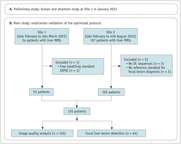
Materials and methods: This retrospective study included 155 consecutive patients (110 male; mean age 62.4 ± 11 years) from two sites who underwent standard liver MRI and additional AMRIDL sequences, specifically DL-accelerated single-shot fast-spin echo (SSFSEDL) and DL-accelerated diffusion-weighted imaging (DWIDL). Additional MRI phantom experiments assessed signal-to-noise ratio (SNR), contrast-to-noise ratio (CNR), and apparent diffusion coefficient (ADC) values. Three reviewers evaluated AMRIDL and AMRISTD protocols for image quality using a five-point Likert scale and identified malignant hepatic lesions. Image quality scores and per-lesion sensitivities were compared between AMRIDL and AMRISTD using the Wilcoxon signed-rank test and logistic regression with generalized estimating equations, respectively.
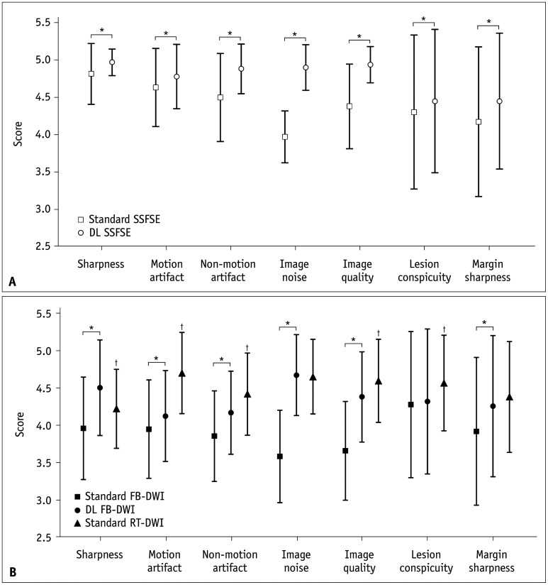
Results: Phantom experiments demonstrated comparable SNR and higher CNR for SSFSEDL compared to SSFSESTD, with similar ADC values for DWIDL and DWISTD. Among the 155 patients, 130 (83.9%) had chronic liver disease or a history of intra- or extrahepatic malignancy. Of 104 malignant focal lesions in 64 patients, 58 (55.8%) were hepatocellular carcinomas (HCCs), 38 (36.5%) were metastases, four (3.8%) were cholangiocarcinomas, and four (3.8%) were lymphomas. The pooled per-lesion sensitivity across three readers was 97.6% for AMRIDL, comparable to 97.6% for AMRISTD. Compared with AMRISTD, AMRIDL demonstrated superior image quality regarding structural sharpness, artifacts, and noise (all P < 0.001) and reduced the average scan time by approximately 50% (2 min 29 sec vs. 4 min 11 sec). In patients with chronic liver disease, AMRIDL achieved a 96.6% per-lesion sensitivity for HCC detection, similar to 96.5% for AMRISTD (P > 0.05).
Conclusion: The AMRIDL protocol offers comparable sensitivity for detecting malignant focal lesions, including HCC while significantly enhancing image quality and reducing scan time by approximately 50% compared to AMRISTD.
期刊介绍:
The inaugural issue of the Korean J Radiol came out in March 2000. Our journal aims to produce and propagate knowledge on radiologic imaging and related sciences.
A unique feature of the articles published in the Journal will be their reflection of global trends in radiology combined with an East-Asian perspective. Geographic differences in disease prevalence will be reflected in the contents of papers, and this will serve to enrich our body of knowledge.
World''s outstanding radiologists from many countries are serving as editorial board of our journal.

 求助内容:
求助内容: 应助结果提醒方式:


