{"title":"A Rapidly Growing Nodule on the Eyebrow of a Pediatric Patient.","authors":"Italo Francesco Aromolo, Michela Brena, Nicola Adriano Monzani, Fabio Caviggioli, Emilio Berti, Donata Micello, Riccardo Cavalli","doi":"10.3390/dermatopathology11040028","DOIUrl":null,"url":null,"abstract":"<p><p>A 11-year-old Caucasian girl presented to our Dermatology Unit with a 2-month history of an erythematous nodule, localized to the medial portion of her left eyebrow, rapidly growing in the two weeks before presentation. The histopathological examination revealed a dermal multi-nodular epithelial neoplasm composed of clear cells, squamous cells, and glandular cells, characterized by cytologic atypia, high mitotic activity, and an infiltrative deep growth pattern. The immunohistochemical profile of the lesion was as follows: CKAE1/AE3+, EMA+, CK8/18+, CK7+, CK19+, AR negative, p63 focally +, Ki67 25%, rare cells GCDFP15+, p53+.</p>","PeriodicalId":42885,"journal":{"name":"Dermatopathology","volume":"11 4","pages":"266-271"},"PeriodicalIF":1.7000,"publicationDate":"2024-09-30","publicationTypes":"Journal Article","fieldsOfStudy":null,"isOpenAccess":false,"openAccessPdf":"https://www.ncbi.nlm.nih.gov/pmc/articles/PMC11503340/pdf/","citationCount":"0","resultStr":null,"platform":"Semanticscholar","paperid":null,"PeriodicalName":"Dermatopathology","FirstCategoryId":"1085","ListUrlMain":"https://doi.org/10.3390/dermatopathology11040028","RegionNum":0,"RegionCategory":null,"ArticlePicture":[],"TitleCN":null,"AbstractTextCN":null,"PMCID":null,"EPubDate":"","PubModel":"","JCR":"Q3","JCRName":"DERMATOLOGY","Score":null,"Total":0}
引用次数: 0
Abstract
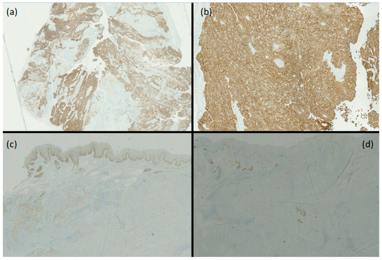
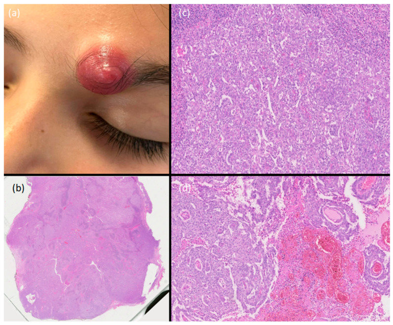
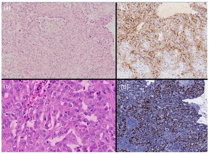
A 11-year-old Caucasian girl presented to our Dermatology Unit with a 2-month history of an erythematous nodule, localized to the medial portion of her left eyebrow, rapidly growing in the two weeks before presentation. The histopathological examination revealed a dermal multi-nodular epithelial neoplasm composed of clear cells, squamous cells, and glandular cells, characterized by cytologic atypia, high mitotic activity, and an infiltrative deep growth pattern. The immunohistochemical profile of the lesion was as follows: CKAE1/AE3+, EMA+, CK8/18+, CK7+, CK19+, AR negative, p63 focally +, Ki67 25%, rare cells GCDFP15+, p53+.

 求助内容:
求助内容: 应助结果提醒方式:


