Clinical Efficacy of Ultrasound-guided High-intensity Focused Ultrasound Ablation for Treating Breast Fibroadenoma of Different Sizes: A Retrospective Study.
Xiuying Wu, Lei Yang, Zi Li, Heng Yin, Wenzhi Chen, Cai Zhang
{"title":"Clinical Efficacy of Ultrasound-guided High-intensity Focused Ultrasound Ablation for Treating Breast Fibroadenoma of Different Sizes: A Retrospective Study.","authors":"Xiuying Wu, Lei Yang, Zi Li, Heng Yin, Wenzhi Chen, Cai Zhang","doi":"10.4103/gmit.GMIT-D-24-00035","DOIUrl":null,"url":null,"abstract":"<p><strong>Objectives: </strong>The aim of this study was to assess the clinical outcomes of ultrasound (US)-guided high-intensity focused ultrasound (HIFU) in patients with breast fibroadenoma (FA) of different sizes.</p><p><strong>Materials and methods: </strong>A total of 88 patients with 245 lesions diagnosed with FA by core needle biopsy from January 2021 to November 2023 were included in this study. US-guided HIFU was performed under local anesthesia. Baseline and treatment characteristics were recorded and analyzed. FAs were divided into three groups according to the longest diameter for further analysis. After the treatment, follow-up with volume evaluation and physical examination was performed at 3, 6, and 12 months.</p><p><strong>Results: </strong>There were 56 FAs ≤10 mm (group 1), 144 FAs with a diameter of 10-20 mm (Group 2), and 45 FAs of 20-30 mm (Group 3). The sonication time of the three groups was 22.5 s, 45.0 s, and 83.0 s (<i>P</i> < 0.05). Based on contrast-enhanced ultrasound evaluation, the median nonperfused volume ratio of the three groups was 74.1%, 87.6%, and 79.2% (<i>P</i> > 0.05), respectively. The volume reduction rates (VRR) of the three groups were 47.3%, 77.0%, and 82.0% at 12 months after HIFU, showing statistical differences. All patients were tolerated well and there were no adverse events after HIFU.</p><p><strong>Conclusion: </strong>The current evidence indicated HIFU was effective and safe in treating breast FA of different sizes, and the VRR of FA >1 cm at 12 months post-HIFU was greater than that of FA <1 cm.</p>","PeriodicalId":45272,"journal":{"name":"Gynecology and Minimally Invasive Therapy-GMIT","volume":"14 1","pages":"72-80"},"PeriodicalIF":1.7000,"publicationDate":"2025-02-27","publicationTypes":"Journal Article","fieldsOfStudy":null,"isOpenAccess":false,"openAccessPdf":"https://www.ncbi.nlm.nih.gov/pmc/articles/PMC11936401/pdf/","citationCount":"0","resultStr":null,"platform":"Semanticscholar","paperid":null,"PeriodicalName":"Gynecology and Minimally Invasive Therapy-GMIT","FirstCategoryId":"1085","ListUrlMain":"https://doi.org/10.4103/gmit.GMIT-D-24-00035","RegionNum":0,"RegionCategory":null,"ArticlePicture":[],"TitleCN":null,"AbstractTextCN":null,"PMCID":null,"EPubDate":"2025/1/1 0:00:00","PubModel":"eCollection","JCR":"Q3","JCRName":"OBSTETRICS & GYNECOLOGY","Score":null,"Total":0}
引用次数: 0
Abstract
Objectives: The aim of this study was to assess the clinical outcomes of ultrasound (US)-guided high-intensity focused ultrasound (HIFU) in patients with breast fibroadenoma (FA) of different sizes.
Materials and methods: A total of 88 patients with 245 lesions diagnosed with FA by core needle biopsy from January 2021 to November 2023 were included in this study. US-guided HIFU was performed under local anesthesia. Baseline and treatment characteristics were recorded and analyzed. FAs were divided into three groups according to the longest diameter for further analysis. After the treatment, follow-up with volume evaluation and physical examination was performed at 3, 6, and 12 months.
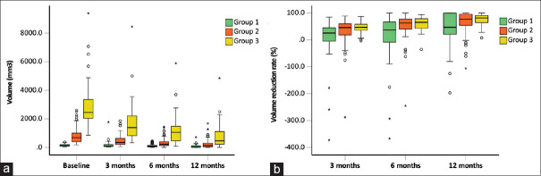
Results: There were 56 FAs ≤10 mm (group 1), 144 FAs with a diameter of 10-20 mm (Group 2), and 45 FAs of 20-30 mm (Group 3). The sonication time of the three groups was 22.5 s, 45.0 s, and 83.0 s (P < 0.05). Based on contrast-enhanced ultrasound evaluation, the median nonperfused volume ratio of the three groups was 74.1%, 87.6%, and 79.2% (P > 0.05), respectively. The volume reduction rates (VRR) of the three groups were 47.3%, 77.0%, and 82.0% at 12 months after HIFU, showing statistical differences. All patients were tolerated well and there were no adverse events after HIFU.
Conclusion: The current evidence indicated HIFU was effective and safe in treating breast FA of different sizes, and the VRR of FA >1 cm at 12 months post-HIFU was greater than that of FA <1 cm.

 求助内容:
求助内容: 应助结果提醒方式:


