Benard Ohene-Botwe, Samuel Anim-Sampong, Robert Saizi
{"title":"Comparison of Anatomical and Indication-Based Diagnostic Reference Levels (DRLs) in Head CT Imaging: Implications for Radiation Dose Management.","authors":"Benard Ohene-Botwe, Samuel Anim-Sampong, Robert Saizi","doi":"10.1155/ijbi/6464273","DOIUrl":null,"url":null,"abstract":"<p><p><b>Introduction:</b> Many diagnostic reference levels (DRLs) in computed tomography (CT) imaging are based mainly on anatomical locations and often overlook variations in radiation exposure due to different clinical indications. While indication-based DRLs, derived from dose descriptors like volume-weighted CT dose index (CTDI<sub>vol</sub>) and dose length product (DLP), are recommended for optimising patient radiation exposure, many studies still use anatomical-based DRL values. This study is aimed at quantifying the differences between anatomical and indication-based DRL values in head CT imaging and assessing its implications for radiation dose management. This will support the narrative when explaining the distinction between indication-based DRLs and anatomical DRLs for patients' dose management. <b>Methods:</b> Employing a retrospective quantitative study design, we developed and compared anatomical and common indication-based DRL values using a dataset of head CT scans with similar characteristics. The indications included in the study were brain tumor/intracranial space-occupying lesion (ISOL), head injury/trauma, stroke, and anatomical examinations. Data analysis was conducted using SPSS Version 29. <b>Results:</b> The findings suggest that using anatomical-based DLP DRL values for CT head examinations leads to underestimations in the median, 25th percentile, and 75th percentile values of head injury/trauma by 20.2%, 30.0%, and 14.5% in single-phase CT head procedures. Conversely, for the entire examination, using anatomical-based DLP DRL as a benchmark for CT stroke DRL overestimates median, 25th percentile, and 75th percentile values by 18.3%, 23.9%, and 13.5%. Brain tumor/ISOL DL<i>P</i> values are underestimated by 62.6%, 60.4%, and 71.8%, respectively. <b>Conclusion:</b>The study highlights that using anatomical DLP DRL values for specific indications in head CT scans can lead to underestimated or overestimated DL<i>P</i> values, making them less reliable for radiation management compared to indication-based DRLs. Therefore, it is imperative to promote the establishment and use of indication-based DRLs for more accurate dose management in CT imaging.</p>","PeriodicalId":47063,"journal":{"name":"International Journal of Biomedical Imaging","volume":"2025 ","pages":"6464273"},"PeriodicalIF":1.3000,"publicationDate":"2025-03-19","publicationTypes":"Journal Article","fieldsOfStudy":null,"isOpenAccess":false,"openAccessPdf":"https://www.ncbi.nlm.nih.gov/pmc/articles/PMC11944678/pdf/","citationCount":"0","resultStr":null,"platform":"Semanticscholar","paperid":null,"PeriodicalName":"International Journal of Biomedical Imaging","FirstCategoryId":"1085","ListUrlMain":"https://doi.org/10.1155/ijbi/6464273","RegionNum":0,"RegionCategory":null,"ArticlePicture":[],"TitleCN":null,"AbstractTextCN":null,"PMCID":null,"EPubDate":"2025/1/1 0:00:00","PubModel":"eCollection","JCR":"Q2","JCRName":"ENGINEERING, BIOMEDICAL","Score":null,"Total":0}
引用次数: 0
Abstract
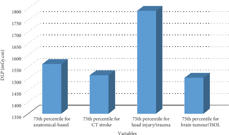
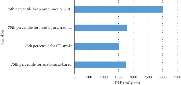
Introduction: Many diagnostic reference levels (DRLs) in computed tomography (CT) imaging are based mainly on anatomical locations and often overlook variations in radiation exposure due to different clinical indications. While indication-based DRLs, derived from dose descriptors like volume-weighted CT dose index (CTDIvol) and dose length product (DLP), are recommended for optimising patient radiation exposure, many studies still use anatomical-based DRL values. This study is aimed at quantifying the differences between anatomical and indication-based DRL values in head CT imaging and assessing its implications for radiation dose management. This will support the narrative when explaining the distinction between indication-based DRLs and anatomical DRLs for patients' dose management. Methods: Employing a retrospective quantitative study design, we developed and compared anatomical and common indication-based DRL values using a dataset of head CT scans with similar characteristics. The indications included in the study were brain tumor/intracranial space-occupying lesion (ISOL), head injury/trauma, stroke, and anatomical examinations. Data analysis was conducted using SPSS Version 29. Results: The findings suggest that using anatomical-based DLP DRL values for CT head examinations leads to underestimations in the median, 25th percentile, and 75th percentile values of head injury/trauma by 20.2%, 30.0%, and 14.5% in single-phase CT head procedures. Conversely, for the entire examination, using anatomical-based DLP DRL as a benchmark for CT stroke DRL overestimates median, 25th percentile, and 75th percentile values by 18.3%, 23.9%, and 13.5%. Brain tumor/ISOL DLP values are underestimated by 62.6%, 60.4%, and 71.8%, respectively. Conclusion:The study highlights that using anatomical DLP DRL values for specific indications in head CT scans can lead to underestimated or overestimated DLP values, making them less reliable for radiation management compared to indication-based DRLs. Therefore, it is imperative to promote the establishment and use of indication-based DRLs for more accurate dose management in CT imaging.
期刊介绍:
The International Journal of Biomedical Imaging is managed by a board of editors comprising internationally renowned active researchers. The journal is freely accessible online and also offered for purchase in print format. It employs a web-based review system to ensure swift turnaround times while maintaining high standards. In addition to regular issues, special issues are organized by guest editors. The subject areas covered include (but are not limited to):
Digital radiography and tomosynthesis
X-ray computed tomography (CT)
Magnetic resonance imaging (MRI)
Single photon emission computed tomography (SPECT)
Positron emission tomography (PET)
Ultrasound imaging
Diffuse optical tomography, coherence, fluorescence, bioluminescence tomography, impedance tomography
Neutron imaging for biomedical applications
Magnetic and optical spectroscopy, and optical biopsy
Optical, electron, scanning tunneling/atomic force microscopy
Small animal imaging
Functional, cellular, and molecular imaging
Imaging assays for screening and molecular analysis
Microarray image analysis and bioinformatics
Emerging biomedical imaging techniques
Imaging modality fusion
Biomedical imaging instrumentation
Biomedical image processing, pattern recognition, and analysis
Biomedical image visualization, compression, transmission, and storage
Imaging and modeling related to systems biology and systems biomedicine
Applied mathematics, applied physics, and chemistry related to biomedical imaging
Grid-enabling technology for biomedical imaging and informatics

 求助内容:
求助内容: 应助结果提醒方式:


