Zeynep Sahin, Deniz Ozkan Vardar, Ekin Erdogmus, Semih Calamak, Belma Koçer Gumusel
{"title":"Monomer release, cytotoxicity, and surface roughness of temporary fixed prosthetic materials produced by digital and conventional methods.","authors":"Zeynep Sahin, Deniz Ozkan Vardar, Ekin Erdogmus, Semih Calamak, Belma Koçer Gumusel","doi":"10.1007/s10266-025-01091-8","DOIUrl":null,"url":null,"abstract":"<p><p>This study compared surface roughness, monomer release, and, cytotoxicity of temporary fixed prosthetic materials manufactured using the conventional, CAD/CAM milling and 3D printing methods. Disc-shaped samples (2 mm height, 5 mm diameter) were prepared from four materials [polyethyl methacrylate/polymethyl methacrylate (Dentalon Plus-DP), bis-acrylic composite resin (Protemp 4-PT), polymethyl methacrylate CAD/CAM disc (On Dent), and methacrylate-based resin (QuraCROWN Temp)]. Surface roughness was measured with a profilometer; scanning electron microscopy (SEM) was used for surface characterization. Following 24, 72, and 120 h of artificial saliva incubation for the samples, the obtained extracts were evaluated for cytotoxicity by performing 3-(4,5-dimethylthiazol-2-yl)-2,5-diphenyltetrazolium bromide (MTT) test in the mouse fibroblast cell. Monomer release from the test samples was analyzed by High‑Performance Liquid Chromatography. Attenuated Total Reflectance Fourier Transform Infrared Spectroscopy (ATR-FTIR) was performed to evaluate the chemical composition of artificial saliva extracts. Cell viability was assessed by one-way ANOVA, and surface roughness by Kruskal-Wallis and Mann-Whitney U tests. No monomer was detected in artificial saliva for any materials. The FTIR spectroscopy of the extracts did not show any peaks corresponding to these monomer or polymer structures, indicating that no residual monomer or polymer was released into the artificial saliva after exposure to artificial saliva. 3D-printed materials were significantly more cytotoxic than the other three test materials at all time points and dilutions (p < 0.05). The highest cell viability rates were detected in CAD/CAM milling (99.43 ± 3.79) at 24 h and PT materials (100.47 ± 5.31) at 72 h for 1:8 dilution. At 1:4 dilution, except for the DP-3D printing test groups, the other groups show similar cell viability rates with the control group (p > 0.05). Digitally manufactured materials had lower roughness than conventionally produced ones (p < 0.05). CAD/CAM milling and PT materials were the most biocompatible, while 3D-printed material was found to be cytotoxic. CAD/CAM milling and PT materials may offer safe and effective options for temporary prosthetic restorations. Although DP showed acceptable results, it was less effective than CAD/CAM milling and PT materials. Due to their cytotoxicity, 3D-printed materials require further investigation before clinical use.</p>","PeriodicalId":19390,"journal":{"name":"Odontology","volume":" ","pages":"1643-1658"},"PeriodicalIF":2.4000,"publicationDate":"2025-10-01","publicationTypes":"Journal Article","fieldsOfStudy":null,"isOpenAccess":false,"openAccessPdf":"https://www.ncbi.nlm.nih.gov/pmc/articles/PMC12450830/pdf/","citationCount":"0","resultStr":null,"platform":"Semanticscholar","paperid":null,"PeriodicalName":"Odontology","FirstCategoryId":"3","ListUrlMain":"https://doi.org/10.1007/s10266-025-01091-8","RegionNum":3,"RegionCategory":"医学","ArticlePicture":[],"TitleCN":null,"AbstractTextCN":null,"PMCID":null,"EPubDate":"2025/3/26 0:00:00","PubModel":"Epub","JCR":"Q2","JCRName":"DENTISTRY, ORAL SURGERY & MEDICINE","Score":null,"Total":0}
引用次数: 0
Abstract
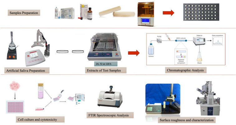
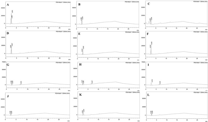
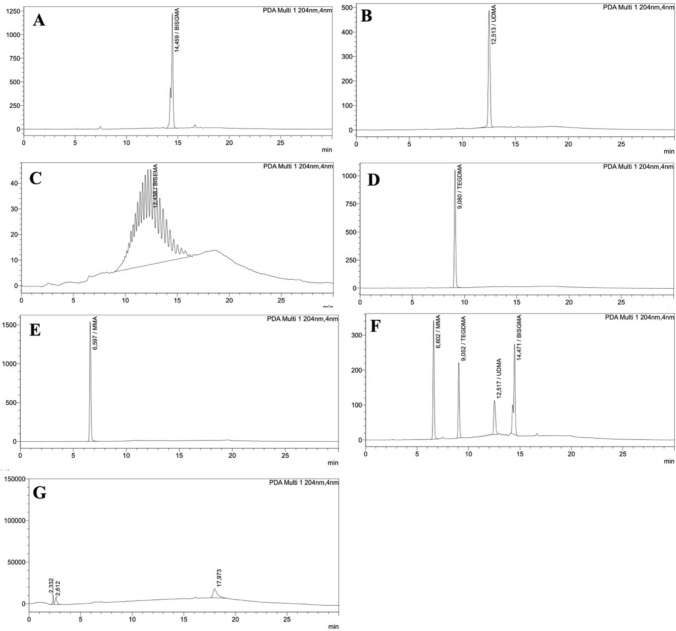
This study compared surface roughness, monomer release, and, cytotoxicity of temporary fixed prosthetic materials manufactured using the conventional, CAD/CAM milling and 3D printing methods. Disc-shaped samples (2 mm height, 5 mm diameter) were prepared from four materials [polyethyl methacrylate/polymethyl methacrylate (Dentalon Plus-DP), bis-acrylic composite resin (Protemp 4-PT), polymethyl methacrylate CAD/CAM disc (On Dent), and methacrylate-based resin (QuraCROWN Temp)]. Surface roughness was measured with a profilometer; scanning electron microscopy (SEM) was used for surface characterization. Following 24, 72, and 120 h of artificial saliva incubation for the samples, the obtained extracts were evaluated for cytotoxicity by performing 3-(4,5-dimethylthiazol-2-yl)-2,5-diphenyltetrazolium bromide (MTT) test in the mouse fibroblast cell. Monomer release from the test samples was analyzed by High‑Performance Liquid Chromatography. Attenuated Total Reflectance Fourier Transform Infrared Spectroscopy (ATR-FTIR) was performed to evaluate the chemical composition of artificial saliva extracts. Cell viability was assessed by one-way ANOVA, and surface roughness by Kruskal-Wallis and Mann-Whitney U tests. No monomer was detected in artificial saliva for any materials. The FTIR spectroscopy of the extracts did not show any peaks corresponding to these monomer or polymer structures, indicating that no residual monomer or polymer was released into the artificial saliva after exposure to artificial saliva. 3D-printed materials were significantly more cytotoxic than the other three test materials at all time points and dilutions (p < 0.05). The highest cell viability rates were detected in CAD/CAM milling (99.43 ± 3.79) at 24 h and PT materials (100.47 ± 5.31) at 72 h for 1:8 dilution. At 1:4 dilution, except for the DP-3D printing test groups, the other groups show similar cell viability rates with the control group (p > 0.05). Digitally manufactured materials had lower roughness than conventionally produced ones (p < 0.05). CAD/CAM milling and PT materials were the most biocompatible, while 3D-printed material was found to be cytotoxic. CAD/CAM milling and PT materials may offer safe and effective options for temporary prosthetic restorations. Although DP showed acceptable results, it was less effective than CAD/CAM milling and PT materials. Due to their cytotoxicity, 3D-printed materials require further investigation before clinical use.
期刊介绍:
The Journal Odontology covers all disciplines involved in the fields of dentistry and craniofacial research, including molecular studies related to oral health and disease. Peer-reviewed articles cover topics ranging from research on human dental pulp, to comparisons of analgesics in surgery, to analysis of biofilm properties of dental plaque.
 求助内容:
求助内容: 应助结果提醒方式:


