Doriane Hazart, Marwa Moulzir, Brigitte Delhomme, Martin Oheim, Clément Ricard
{"title":"Imaging the enteric nervous system.","authors":"Doriane Hazart, Marwa Moulzir, Brigitte Delhomme, Martin Oheim, Clément Ricard","doi":"10.3389/fnana.2025.1532900","DOIUrl":null,"url":null,"abstract":"<p><p>The enteric nervous system (ENS) has garnered increasing scientific interest due to its pivotal role in digestive processes and its involvement in various gastrointestinal and central nervous system (CNS) disorders, including Crohn's disease, Parkinson's disease, and autism. Despite its significance, the ENS remains relatively underexplored by neurobiologists, primarily because its structure and function are less understood compared to the CNS. This review examines both pioneering methodologies that initially revealed the intricate layered structure of the ENS and recent advancements in studying its three-dimensional (3-D) organization, both in fixed samples and at a functional level, <i>ex-vivo</i> or <i>in-vivo</i>. Traditionally, imaging the ENS relied on histological techniques involving sequential tissue sectioning, staining, and microscopic imaging of single sections. However, this method has limitations representing the full complexity of the ENS's 3-D meshwork, which led to the development of more intact preparations, such as whole-mount preparation, as well as the use of volume imaging techniques. Advancements in 3-D imaging, particularly methods like spinning-disk confocal, 2-photon, and light-sheet microscopies, combined with tissue-clearing techniques, have revolutionized our understanding of the ENS's fine structure. These approaches offer detailed views of its cellular architecture, including interactions among various cell types, blood vessels, and lymphatic vessels. They have also enhanced our comprehension of ENS-related pathologies, such as inflammatory bowel disease, Hirschsprung's disease (HSCR), and the ENS's involvement in neurodegenerative disorders like Parkinson's (PD) and Alzheimer's diseases (AD). More recently, 2-photon or confocal <i>in-vivo</i> imaging, combined with transgenic approaches for calcium imaging, or confocal laser endomicroscopy, have opened new avenues for functional studies of the ENS. These methods enable real-time observation of enteric neuronal and glial activity and their interactions. While routinely used in CNS studies, their application to understanding local circuits and signals in the ENS is relatively recent and presents unique challenges, such as accommodating peristaltic movements. Advancements in 3-D <i>in-vivo</i> functional imaging are expected to significantly deepen our understanding of the ENS and its roles in gastrointestinal and neurological diseases, potentially leading to improved diagnostic and therapeutic strategies.</p>","PeriodicalId":12572,"journal":{"name":"Frontiers in Neuroanatomy","volume":"19 ","pages":"1532900"},"PeriodicalIF":2.3000,"publicationDate":"2025-03-12","publicationTypes":"Journal Article","fieldsOfStudy":null,"isOpenAccess":false,"openAccessPdf":"https://www.ncbi.nlm.nih.gov/pmc/articles/PMC11937143/pdf/","citationCount":"0","resultStr":null,"platform":"Semanticscholar","paperid":null,"PeriodicalName":"Frontiers in Neuroanatomy","FirstCategoryId":"3","ListUrlMain":"https://doi.org/10.3389/fnana.2025.1532900","RegionNum":4,"RegionCategory":"医学","ArticlePicture":[],"TitleCN":null,"AbstractTextCN":null,"PMCID":null,"EPubDate":"2025/1/1 0:00:00","PubModel":"eCollection","JCR":"Q1","JCRName":"ANATOMY & MORPHOLOGY","Score":null,"Total":0}
引用次数: 0
Abstract
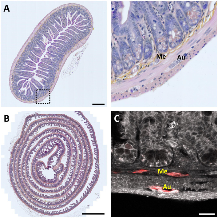
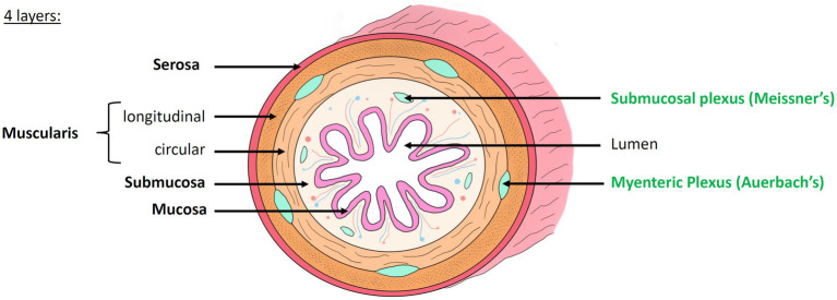
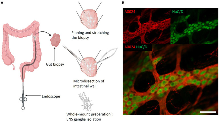
The enteric nervous system (ENS) has garnered increasing scientific interest due to its pivotal role in digestive processes and its involvement in various gastrointestinal and central nervous system (CNS) disorders, including Crohn's disease, Parkinson's disease, and autism. Despite its significance, the ENS remains relatively underexplored by neurobiologists, primarily because its structure and function are less understood compared to the CNS. This review examines both pioneering methodologies that initially revealed the intricate layered structure of the ENS and recent advancements in studying its three-dimensional (3-D) organization, both in fixed samples and at a functional level, ex-vivo or in-vivo. Traditionally, imaging the ENS relied on histological techniques involving sequential tissue sectioning, staining, and microscopic imaging of single sections. However, this method has limitations representing the full complexity of the ENS's 3-D meshwork, which led to the development of more intact preparations, such as whole-mount preparation, as well as the use of volume imaging techniques. Advancements in 3-D imaging, particularly methods like spinning-disk confocal, 2-photon, and light-sheet microscopies, combined with tissue-clearing techniques, have revolutionized our understanding of the ENS's fine structure. These approaches offer detailed views of its cellular architecture, including interactions among various cell types, blood vessels, and lymphatic vessels. They have also enhanced our comprehension of ENS-related pathologies, such as inflammatory bowel disease, Hirschsprung's disease (HSCR), and the ENS's involvement in neurodegenerative disorders like Parkinson's (PD) and Alzheimer's diseases (AD). More recently, 2-photon or confocal in-vivo imaging, combined with transgenic approaches for calcium imaging, or confocal laser endomicroscopy, have opened new avenues for functional studies of the ENS. These methods enable real-time observation of enteric neuronal and glial activity and their interactions. While routinely used in CNS studies, their application to understanding local circuits and signals in the ENS is relatively recent and presents unique challenges, such as accommodating peristaltic movements. Advancements in 3-D in-vivo functional imaging are expected to significantly deepen our understanding of the ENS and its roles in gastrointestinal and neurological diseases, potentially leading to improved diagnostic and therapeutic strategies.
期刊介绍:
Frontiers in Neuroanatomy publishes rigorously peer-reviewed research revealing important aspects of the anatomical organization of all nervous systems across all species. Specialty Chief Editor Javier DeFelipe at the Cajal Institute (CSIC) is supported by an outstanding Editorial Board of international experts. This multidisciplinary open-access journal is at the forefront of disseminating and communicating scientific knowledge and impactful discoveries to researchers, academics, clinicians and the public worldwide.
 求助内容:
求助内容: 应助结果提醒方式:


