Jorge Carlos Alberdi, Ignacio Troiano, Miguel Angel Troiano, Patricia Mónica Sanchez, Flávio Rodrigues Ferreira Alves
{"title":"Effective Management of Calcified Root Canals Using Static-guided Access: A Case Series.","authors":"Jorge Carlos Alberdi, Ignacio Troiano, Miguel Angel Troiano, Patricia Mónica Sanchez, Flávio Rodrigues Ferreira Alves","doi":"10.14744/eej.2024.66588","DOIUrl":null,"url":null,"abstract":"<p><p>Root canal calcification poses a substantial challenge in endodontic practice and may lead to treatment failure. The difficulty lies in accessing, penetrating, and negotiating these canals. This article reports on a series of calcified root canals successfully treated using static-guided endodontics. Eleven cases of calcified root canals were treated by the same endodontist using static-guided endodontics. The sample encompassed four tooth types including a premolar with two canals, effectively managed using two templates (drill guides). Guided endodontic treatment was based on evidence of calcified root canals diagnosed with clinical, radiographic, and cone beam computed tomography (CBCT) findings. A high-resolution CBCT and an intraoral scan were used for virtual cavity planning. The CBCT and intraoral scan were superimposed, and virtual sleeves were accurately placed to avoid drilling deviation. Templates were fabricated and fitted, and guided access was conducted with low-speed drilling, monitored with intraoperative radiographs. Canals were negotiated with K-files, and prepared with Wave One Gold system, using 2.5% NaOCl as irrigant. In all cases, virtually planned guided coronal and root canal access allied to the 3D printed templates allowed canal location through obliterated pulp spaces with a conservative access approach and without accidents. The cases demonstrated that static-guided endodontics is a safe accurate treatment approach to access calcified canals, reducing working time, minimizing removal of tooth structure, and decreasing the risk of iatrogenic damage. (EEJ-2024-07-106).</p>","PeriodicalId":11860,"journal":{"name":"European Endodontic Journal","volume":"10 1","pages":"73-82"},"PeriodicalIF":2.0000,"publicationDate":"2025-01-01","publicationTypes":"Journal Article","fieldsOfStudy":null,"isOpenAccess":false,"openAccessPdf":"https://www.ncbi.nlm.nih.gov/pmc/articles/PMC11971709/pdf/","citationCount":"0","resultStr":null,"platform":"Semanticscholar","paperid":null,"PeriodicalName":"European Endodontic Journal","FirstCategoryId":"1085","ListUrlMain":"https://doi.org/10.14744/eej.2024.66588","RegionNum":0,"RegionCategory":null,"ArticlePicture":[],"TitleCN":null,"AbstractTextCN":null,"PMCID":null,"EPubDate":"","PubModel":"","JCR":"Q3","JCRName":"DENTISTRY, ORAL SURGERY & MEDICINE","Score":null,"Total":0}
引用次数: 0
Abstract
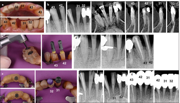
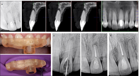
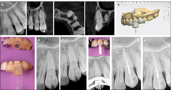
Root canal calcification poses a substantial challenge in endodontic practice and may lead to treatment failure. The difficulty lies in accessing, penetrating, and negotiating these canals. This article reports on a series of calcified root canals successfully treated using static-guided endodontics. Eleven cases of calcified root canals were treated by the same endodontist using static-guided endodontics. The sample encompassed four tooth types including a premolar with two canals, effectively managed using two templates (drill guides). Guided endodontic treatment was based on evidence of calcified root canals diagnosed with clinical, radiographic, and cone beam computed tomography (CBCT) findings. A high-resolution CBCT and an intraoral scan were used for virtual cavity planning. The CBCT and intraoral scan were superimposed, and virtual sleeves were accurately placed to avoid drilling deviation. Templates were fabricated and fitted, and guided access was conducted with low-speed drilling, monitored with intraoperative radiographs. Canals were negotiated with K-files, and prepared with Wave One Gold system, using 2.5% NaOCl as irrigant. In all cases, virtually planned guided coronal and root canal access allied to the 3D printed templates allowed canal location through obliterated pulp spaces with a conservative access approach and without accidents. The cases demonstrated that static-guided endodontics is a safe accurate treatment approach to access calcified canals, reducing working time, minimizing removal of tooth structure, and decreasing the risk of iatrogenic damage. (EEJ-2024-07-106).

 求助内容:
求助内容: 应助结果提醒方式:


