{"title":"Effect of Various Disinfection Protocols on Endodontic Biofilm and Growth Factors Release from Radicular Dentin: An In Vitro Study.","authors":"Abhilasha Gugliani, Sonali Taneja, Devi Charan Shetty, Vidhi Kiran Bhalla","doi":"10.14744/eej.2024.84856","DOIUrl":null,"url":null,"abstract":"<p><strong>Objective: </strong>The aim of this study was to evaluate and compare the effect of various disinfection protocols on bacterial biofilm and subsequent release of growth factors from radicular dentin.</p><p><strong>Methods: </strong>One hundred and ninety two extracted single rooted premolars were obtained and contaminated with E. faecalis biofilm for 21 days. The samples were then divided into three main groups - Group I: Irrigation (I) only, Group II: Calcium hydroxide (CH) placement followed by final irrigation and Group III: Triple Antibiotic paste (TAP) placement followed by final irrigation. Each group was further then divided into four sub-groups according to the final irrigating solution used - Sub group A: Saline, Sub group B: 17% EDTA, Sub group C: 1% phytic acid and Sub group D: 0.2%. chitosan nanoparticles. After treatment, the samples were subjected to colony-forming unit (CFU) analysis to determine bacterial reduction and the release of TGF-β1 and VEGF from the root canals, which was quantified using Enzyme-Linked Immunosorbent Assay (ELISA). The data were analyzed using statistical tests.</p><p><strong>Results: </strong>The maximum reduction in E. faecalis biofilm was observed in Group III (TAP), followed by Group II (CH), and finally Group I (irrigation only). Among the subgroups, the maximum reduction in bacterial biofilm was seen with chitosan nanoparticles, followed by phytic acid, EDTA, and saline. After 24 hours, the highest release of both TGF-β1 and VEGF was observed in the chitosan nanoparticles subgroup, followed by phytic acid, EDTA, and saline. Similar results were seen in the CH and TAP groups.</p><p><strong>Conclusion: </strong>The study concluded that newer irrigating solutions, particularly 0.2% chitosan nanoparticles, showed superior antibacterial activity and better smear layer removal, leading to greater growth factor release from the radicular dentin. The study also highlighted that TAP placement resulted in maximum bacterial reduction, regardless of the final irrigant used. Furthermore, the release of TGF-β1 was significantly higher than VEGF in all groups. (EEJ-2024-03-045).</p>","PeriodicalId":11860,"journal":{"name":"European Endodontic Journal","volume":"10 1","pages":"1-10"},"PeriodicalIF":2.0000,"publicationDate":"2025-01-01","publicationTypes":"Journal Article","fieldsOfStudy":null,"isOpenAccess":false,"openAccessPdf":"https://www.ncbi.nlm.nih.gov/pmc/articles/PMC11971708/pdf/","citationCount":"0","resultStr":null,"platform":"Semanticscholar","paperid":null,"PeriodicalName":"European Endodontic Journal","FirstCategoryId":"1085","ListUrlMain":"https://doi.org/10.14744/eej.2024.84856","RegionNum":0,"RegionCategory":null,"ArticlePicture":[],"TitleCN":null,"AbstractTextCN":null,"PMCID":null,"EPubDate":"","PubModel":"","JCR":"Q3","JCRName":"DENTISTRY, ORAL SURGERY & MEDICINE","Score":null,"Total":0}
引用次数: 0
Abstract
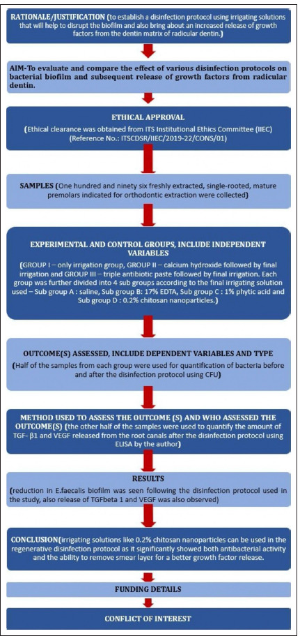
Objective: The aim of this study was to evaluate and compare the effect of various disinfection protocols on bacterial biofilm and subsequent release of growth factors from radicular dentin.
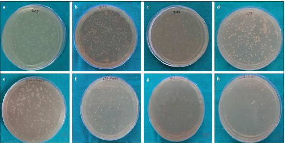
Methods: One hundred and ninety two extracted single rooted premolars were obtained and contaminated with E. faecalis biofilm for 21 days. The samples were then divided into three main groups - Group I: Irrigation (I) only, Group II: Calcium hydroxide (CH) placement followed by final irrigation and Group III: Triple Antibiotic paste (TAP) placement followed by final irrigation. Each group was further then divided into four sub-groups according to the final irrigating solution used - Sub group A: Saline, Sub group B: 17% EDTA, Sub group C: 1% phytic acid and Sub group D: 0.2%. chitosan nanoparticles. After treatment, the samples were subjected to colony-forming unit (CFU) analysis to determine bacterial reduction and the release of TGF-β1 and VEGF from the root canals, which was quantified using Enzyme-Linked Immunosorbent Assay (ELISA). The data were analyzed using statistical tests.
Results: The maximum reduction in E. faecalis biofilm was observed in Group III (TAP), followed by Group II (CH), and finally Group I (irrigation only). Among the subgroups, the maximum reduction in bacterial biofilm was seen with chitosan nanoparticles, followed by phytic acid, EDTA, and saline. After 24 hours, the highest release of both TGF-β1 and VEGF was observed in the chitosan nanoparticles subgroup, followed by phytic acid, EDTA, and saline. Similar results were seen in the CH and TAP groups.
Conclusion: The study concluded that newer irrigating solutions, particularly 0.2% chitosan nanoparticles, showed superior antibacterial activity and better smear layer removal, leading to greater growth factor release from the radicular dentin. The study also highlighted that TAP placement resulted in maximum bacterial reduction, regardless of the final irrigant used. Furthermore, the release of TGF-β1 was significantly higher than VEGF in all groups. (EEJ-2024-03-045).

 求助内容:
求助内容: 应助结果提醒方式:


