{"title":"A Protocol for Void Detection in Root-filled Teeth Using Micro-CT: Ex-vivo.","authors":"Iad Gharib, Ferranti S Wong, Graham Roy Davis","doi":"10.14744/eej.2023.37167","DOIUrl":null,"url":null,"abstract":"<p><strong>Objective: </strong>X-ray microtomography (micro-CT or XMT) has previously been used to measure residual voids in root fillings. However, there is no agreement on a protocol that critically identifies and attempts to solve artefacts inherent to the micro-computed tomography technique. This article aims to describe a protocol for automated detection of voids within root-filled canals taking into account the inherent artefacts, with special interest in the partial volume effect. This is to reduce human errors and increase the accuracy and efficiency of void detection.</p><p><strong>Methods: </strong>Human maxillary premolars (n=33) were shaped, cleaned and root-filled using the cold lateral condensation (CLC) technique. Voids were identified using either individual tomographic slices or the new proposed protocol in which: (1) pre-obturation XMT slices were used to identify the coordinates of the canal space; (2) the post-obturation data sets were aligned to the pre-obturation data sets; (3) the voids were identified as voxels with a grey level below a set threshold after subtraction of pre-obturation from post-obturation data sets. A comparison of the voids from these two methods was made.</p><p><strong>Results: </strong>The visual inspection of slice by slice of the scanned data resulted in full agreement between the tomographic slices and the results gained from the proposed protocol. This confirmed that this protocol provided an automated, effective and accurate method for detecting voids in root-filled canals.</p><p><strong>Conclusion: </strong>The proposed protocol provides an automated method to eliminate inaccuracies from XMT artefacts so that accurate volumetric measurements can be easily obtained. (EEJ-2024-02-031).</p>","PeriodicalId":11860,"journal":{"name":"European Endodontic Journal","volume":"10 1","pages":"11-17"},"PeriodicalIF":2.0000,"publicationDate":"2025-01-01","publicationTypes":"Journal Article","fieldsOfStudy":null,"isOpenAccess":false,"openAccessPdf":"https://www.ncbi.nlm.nih.gov/pmc/articles/PMC11971713/pdf/","citationCount":"0","resultStr":null,"platform":"Semanticscholar","paperid":null,"PeriodicalName":"European Endodontic Journal","FirstCategoryId":"1085","ListUrlMain":"https://doi.org/10.14744/eej.2023.37167","RegionNum":0,"RegionCategory":null,"ArticlePicture":[],"TitleCN":null,"AbstractTextCN":null,"PMCID":null,"EPubDate":"","PubModel":"","JCR":"Q3","JCRName":"DENTISTRY, ORAL SURGERY & MEDICINE","Score":null,"Total":0}
引用次数: 0
Abstract
Objective: X-ray microtomography (micro-CT or XMT) has previously been used to measure residual voids in root fillings. However, there is no agreement on a protocol that critically identifies and attempts to solve artefacts inherent to the micro-computed tomography technique. This article aims to describe a protocol for automated detection of voids within root-filled canals taking into account the inherent artefacts, with special interest in the partial volume effect. This is to reduce human errors and increase the accuracy and efficiency of void detection.
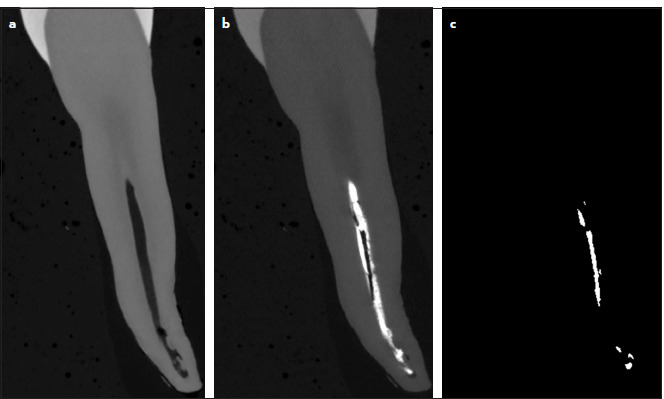
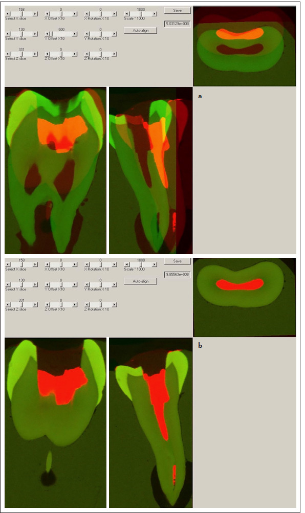
Methods: Human maxillary premolars (n=33) were shaped, cleaned and root-filled using the cold lateral condensation (CLC) technique. Voids were identified using either individual tomographic slices or the new proposed protocol in which: (1) pre-obturation XMT slices were used to identify the coordinates of the canal space; (2) the post-obturation data sets were aligned to the pre-obturation data sets; (3) the voids were identified as voxels with a grey level below a set threshold after subtraction of pre-obturation from post-obturation data sets. A comparison of the voids from these two methods was made.
Results: The visual inspection of slice by slice of the scanned data resulted in full agreement between the tomographic slices and the results gained from the proposed protocol. This confirmed that this protocol provided an automated, effective and accurate method for detecting voids in root-filled canals.
Conclusion: The proposed protocol provides an automated method to eliminate inaccuracies from XMT artefacts so that accurate volumetric measurements can be easily obtained. (EEJ-2024-02-031).

 求助内容:
求助内容: 应助结果提醒方式:


